Japanese
English
- 有料閲覧
- Abstract 文献概要
- 1ページ目 Look Inside
- 参考文献 Reference
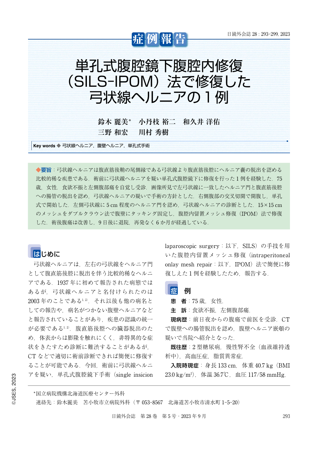
◆要旨:弓状線ヘルニアは腹直筋後鞘の尾側縁である弓状線より腹直筋後腔にヘルニア囊の脱出を認める比較的稀な疾患である.術前に弓状線ヘルニアを疑い単孔式腹腔鏡下に修復を行った1例を経験した.75歳,女性.食欲不振と左側腹部痛を自覚し受診.画像所見で左弓状線に一致したヘルニア門と腹直筋後腔への腸管の脱出を認め,弓状線ヘルニアの疑いで手術の方針とした.右側腹部の交叉切開で開腹し,単孔式で開始した.左側弓状線に5cm程度のヘルニア門を認め,弓状線ヘルニアの診断とした.15×15cmのメッシュをダブルクラウン法で腹壁にタッキング固定し,腹腔内留置メッシュ修復(IPOM)法で修復した.術後腹痛は改善し,9日後に退院,再発なく6か月が経過している.
Arcuate line hernia is a relatively rare condition in which a hernia sac protrudes from the caudal border of the posterior rectus sheath to the dorsal rectus abdominis muscle. We report a case in which an arcuate line hernia was suspected preoperatively and a single incision laparoscopic surgery was performed. A 75-year-old woman presented with anorexia and left-sided abdominal pain. Computed tomography revealed a small bowel protrusion between the rectus abdominis and the posterior rectus sheath from the left arcuate line.The arcuate line hernia was suspected, and surgery was planned. A 2 cm incision was made on the right side of the abdomen, and the operation was started through a single incision laparoscopy. A hernia sac was found in the posterior space of the rectus abdominis muscle in the cephalic direction from the left arcuate line. Tack fixation of 15x15 cm mesh was performed on the abdominal wall using the double crown technique and repaired with an intraperitoneal onlay mesh repair. Postoperatively, abdominal pain improved and the patient was discharged from the hospital 9 days later. No recurrence has occurred in 6 months of follow-up.

Copyright © 2023, JAPAN SOCIETY FOR ENDOSCOPIC SURGERY All rights reserved.


