Japanese
English
- 有料閲覧
- Abstract 文献概要
- 1ページ目 Look Inside
- 参考文献 Reference
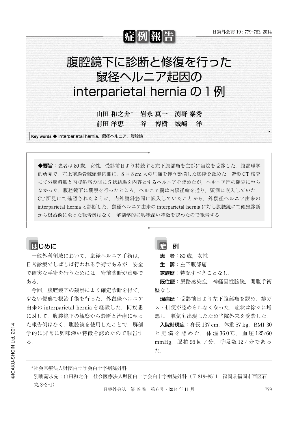
◆要旨:患者は80歳,女性.受診前日より持続する左下腹部痛を主訴に当院を受診した.腹部理学的所見で,左上前腸骨棘頭側内側に,8×8cm大の圧痛を伴う緊満した膨隆を認めた.造影CT検査にて外腹斜筋と内腹斜筋の間にS状結腸を内容とするヘルニアを認めたが,ヘルニア門の確定に至らなかった.腹腔鏡下に観察を行ったところ,ヘルニア囊は内鼠径輪を通り,頭側に嵌入していた.CT所見にて確認されたように,内外腹斜筋間に嵌入していたことから,外鼠径ヘルニア由来のinterparietal herniaと診断した.鼠径ヘルニア由来のinterparietal herniaに対し腹腔鏡にて確定診断から根治術に至った報告例はなく,解剖学的に興味深い特徴を認めたので報告する.
An 80-year-old woman who had no surgical history presented with lower abdominal pain persisting for a day, and a mass that extended cranial medial of the left anterior superior iliac spine of the abdominal wall. Computed tomography(CT) scan revealed that the sigmoid colon was present between the internal and the external oblique muscle. After 5 days of admission, laparoscopic surgery was performed. Laparoscopic examination to identify the hernia orifice revealed that it was the deep inguinal ring and a hernia sac was observed passing through the deep inguinal ring. The hernia sac was extending cranially, and was extended between the external and the internal oblique muscle. Laparoscopic trans-abdominal preperitoneal mesh repair(TAPP) was performed successfully, and no postoperative complication occurred. There have been no previous reports of laparoscopic interparietal hernia repair. This may be the first report, and the strategy seems to be useful for accurate diagnosis and simultaneous treatment with minimal surgical invasiveness.

Copyright © 2014, JAPAN SOCIETY FOR ENDOSCOPIC SURGERY All rights reserved.


