Japanese
English
- 有料閲覧
- Abstract 文献概要
- 1ページ目 Look Inside
- 参考文献 Reference
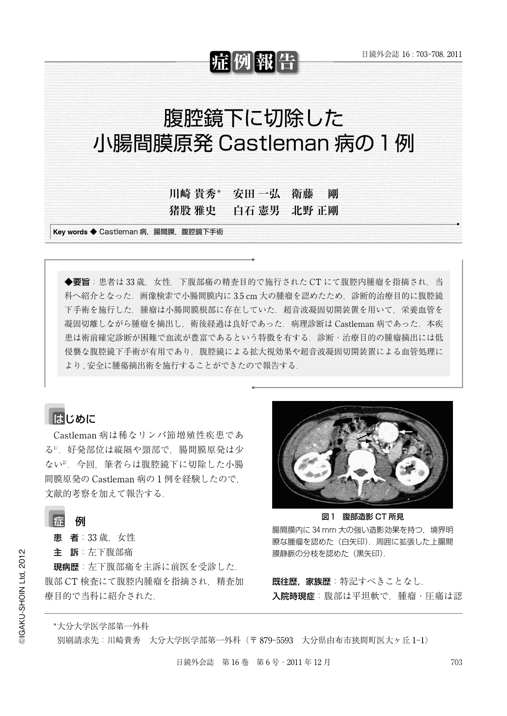
◆要旨:患者は33歳,女性.下腹部痛の精査目的で施行されたCTにて腹腔内腫瘤を指摘され,当科へ紹介となった.画像検索で小腸間膜内に3.5cm大の腫瘤を認めたため,診断的治療目的に腹腔鏡下手術を施行した.腫瘤は小腸間膜根部に存在していた.超音波凝固切開装置を用いて,栄養血管を凝固切離しながら腫瘤を摘出し,術後経過は良好であった.病理診断はCastleman病であった.本疾患は術前確定診断が困難で血流が豊富であるという特徴を有する.診断・治療目的の腫瘤摘出には低侵襲な腹腔鏡下手術が有用であり,腹腔鏡による拡大視効果や超音波凝固切開装置による血管処理により,安全に腫瘍摘出術を施行することができたので報告する.
A 33-year-old woman presented to the hospital complaining of lower abdominal pain. Abdominal CT revealed an intra-abdominal mass, and she was referred to our hospital for treatment. Contrast-enhanced CT and MRI showed a mass measuring 3.5 cm in diameter with heterogeneous enhancement located in the mesentery of the small intestine. We suspected angiosarcoma, gastrointestinal stromal tumor or Castleman's disease, and laparoscopic surgery was performed. A solid tumor was located in the mesentery near the ligament of Treitz and was resected safely with ultrasonic shears. The histopathological diagnosis was unicentric Castleman's disease of hyaline vascular type. Castleman's disease is difficult to diagnose preoperatively and is characterized by its rich blood supply. Laparoscopic surgery is useful for diagnostic treatment because of its minimal invasiveness. We report herein a patient with mesenteric Castleman's disease that was safely resected by laparoscopic surgery.

Copyright © 2011, JAPAN SOCIETY FOR ENDOSCOPIC SURGERY All rights reserved.


