Japanese
English
- 有料閲覧
- Abstract 文献概要
- 1ページ目 Look Inside
I.はじめに
内耳の先天奇形を診断することは今日に至っても困難なことが多い。これまでにも側頭骨病変における断層撮影の有用性については種々詳述されている1〜5)が,日常臨床では読影に苦しむ症例も少なくない。しかし,近年めざましく進歩したCTを併用することによって,側頭骨病変をかなり細かく観察できるようになってきた6〜9)。
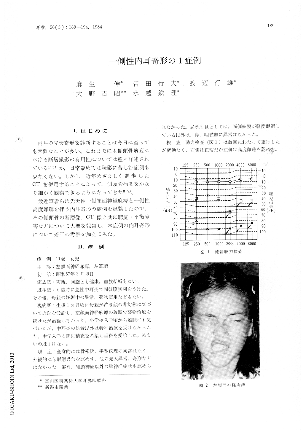
最近筆者らは先天性一側顔面神経麻痺と一側性高度難聴を伴う内耳奇形の症例を経験したので,その側頭骨の断層像,CT像と共に聴覚・平衡障害などについて大要を報告し,本症例の内耳奇形について若干の考察を加えてみた。
Polytomography and CT-scanning of the temporal bone and neuro-otological findings in a 11-year-old girl with left hearing impairment and facial paralysis showed the characteristic features of an unilateral inner ear anomaly. In the affected side the cochlea, vestibule and semicircular canal were intact, but the internal acoustic meatus was narrow because of abnormal bony proliferation. It was suspected that this narrowing of the canal resulted in severe hearing impairment and incomplete facial paralysis. CT-scanning is an important technique because it reveals a fine contour of the inner ear anomaly.

Copyright © 1984, Igaku-Shoin Ltd. All rights reserved.


