Japanese
English
- 有料閲覧
- Abstract 文献概要
- 1ページ目 Look Inside
I.はじめに
鼻副鼻腔領域の真菌症がはじめて報告されたのは,国外においては1791年のPlaignaud1),Hernu2)の各症例,国内では1942年の村田3)の2症例とされているが,近年報告例は増加しつつあり,一側性の副鼻腔陰影をみた際には,上顎癌や歯性上顎洞炎などとともに鑑別すべき疾患の1つとして列挙せねばならないものとなってきている。従来,鼻副鼻腔真菌症は,稀有例としての症例報告の他に,乾酪性副鼻腔炎の原因である点が強調されたり4,5),臨床的に破壊型(侵襲型)と非侵襲型に分類が可能6,7)で,前者は上顎癌と鑑別が困難であったという面からとりあげられている。さらにムコール症の場合は致命率がきわめて高いことが強調されている。
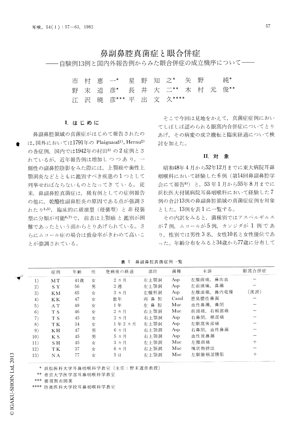
そこで今回は見地をかえて,真菌症症例においてしぽしば認められる眼窩内合併症についてとりあげ,その病変の成立機転と臨床経過について検討を加えた。
Thirteen patients with mycotic infection of the nose and the paranasal sinuses are presented. A review of 438 reported cases in the literature concerning the sinonasal mycosis is also presented in view of the orbital complications.
Most of the orbital complications in aspergillosis are due to a mass lesion in the orbit following the bony destruction of the orbital wall by aspergilloma. Phycomycosis usually starts in the nose and the maxillary sinuses (this stage is considered to be a subclinical one) and spreads to the ethmoid cells and thence to the orbit via the ethmoid vessels. The fungus commonly ascends to the orbital apex and then through the cavernous sinus to invade and obstruct the internal carotid artery in early stage, which leads to variety of orbital symptoms. Exceptional clinical course may exist in both mycoses.
Diagnosis of phycomycosis should be made early in the subclinical stage. The importance of the exploratory antrotomy in the patients with unilateral antral opacity should be emphasized again in this point of view.

Copyright © 1982, Igaku-Shoin Ltd. All rights reserved.


