Japanese
English
- 有料閲覧
- Abstract 文献概要
- 1ページ目 Look Inside
1.はじめに
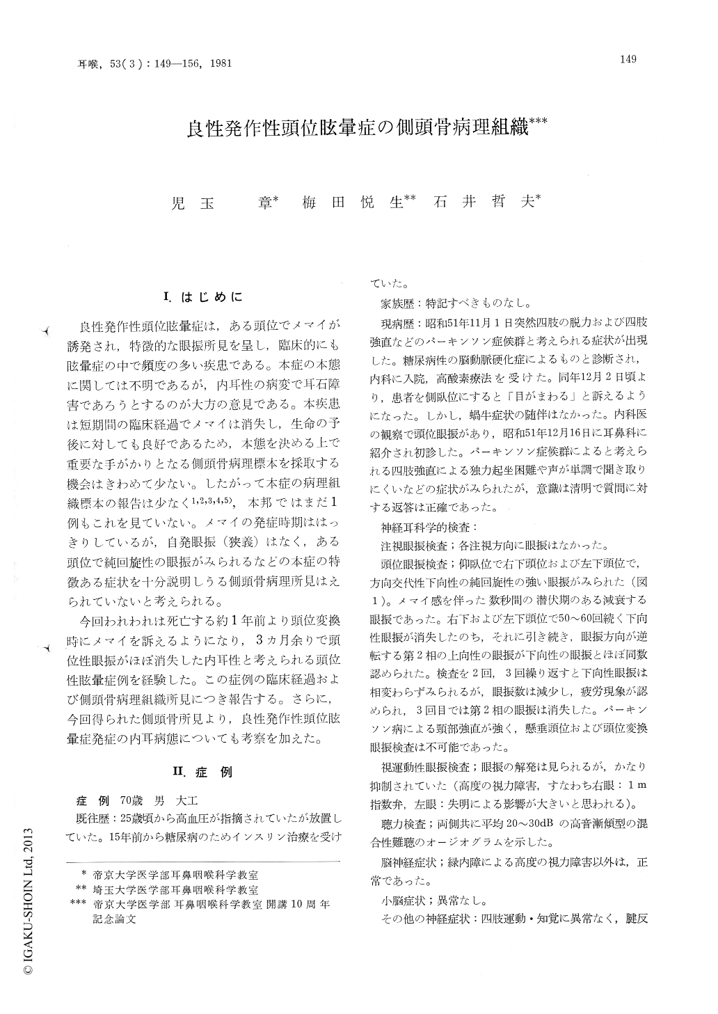
良性発作性頭位眩暈症は,ある頭位でメマイが誘発され,特徴的な眼振所見を呈し,臨床的にも眩暈症の中で頻度の多い疾患である。本症の本態に関しては不明であるが,内耳性の病変で耳石障害であろうとするのが大方の意見である。本疾患は短期間の臨床経過でメマイは消失し,生命の予後に対しても良好であるため,本態を決める上で重要な手がかりとなる側頭骨病理標本を採取する機会はきわめて少ない。したがって本症の病理組織標本の報告は少なく1,2,3,4,5),本邦ではまだ1例もこれを見ていない。メマイの発症時期ははっきりしているが,自発眼振(狭義)はなく,ある頭位で純回旋性の眼振がみられるなどの本症の特徴ある症状を十分説明しうる側頭骨病理所見はえられていないと考えられる。
今回われわれは死亡する約1年前より頭位変換時にメマイを訴えるようになり,3カ月余りで頭位性眼振がほぼ消失した内耳性と考えられる頭位性眩暈症例を経験した。この症例の臨床経過および側頭骨病理組織所見につき報告する。さらに,今回得られた側頭骨所見より,良性発作性頭位眩暈症発症の内耳病態についても考察を加えた。
Benign paroxysmal positional vertigo (BPPN) is one of the common vertiginous diseases, however, there is a lack of report of the temporal bone study of BPPN. The reports, available at present, do not convincingly explain characteristic positional or positioning nystagmus of this disease.
In this paper the histopathological findings of the temporal bones of a patient who exhibited typical positional vertigo of benign paroxysmal type are reported. The main pathological findings were separation of otoconia from the otolithic membrane of the utricular and saccular macula, besides basophilic deposit on the cupula of the posterior semicircular canal. There was no diminution of the sensory cells and nerve fibers in each vestibular endorgans.
These temporal bone findings indicate that vestibular disorder induced by positional or positioning provocation in BPPN of this case was caused by removal of otoconia from the otolithic macula of both ears.

Copyright © 1981, Igaku-Shoin Ltd. All rights reserved.


