Japanese
English
- 有料閲覧
- Abstract 文献概要
- 1ページ目 Look Inside
I.はじめに
下眼窩裂は,眼窩底を内後方より外前方に斜めに走行する裂孔である。その長さは約3cm前後であり1〜3),幅は前端部の広い所では約5mmで,ほぼ中央の狭い所では約2mmである4)。下眼窩裂の上縁の大部分は蝶形骨大翼眼窩面の下縁である眼窩下稜(crista infraorbitalis)よりなる。下眼窩裂下縁は上顎洞後上壁,すなわら上顎骨眼窩面と側頭面とがなす移行縁である5,6)(下眼窩裂前端膨大部では,頬骨が参入する場合がある1)。下縁のほぼ中央には,眼窩下溝が存在し,その内部を眼窩下神経,眼窩下動脈などが通過している7)。下眼窩裂後下方では翼口蓋窩と通じ,後側方では側頭下窩に通じている8,9),下眼窩裂の上内方には,視神経管と上眼窩裂が存在する。このように下眼窩裂は解剖学的に極めて重要な位置を占めるにもかかわらず,その読影に適したX線撮影法は,十分に普及して利用されることが少ない印象がある。
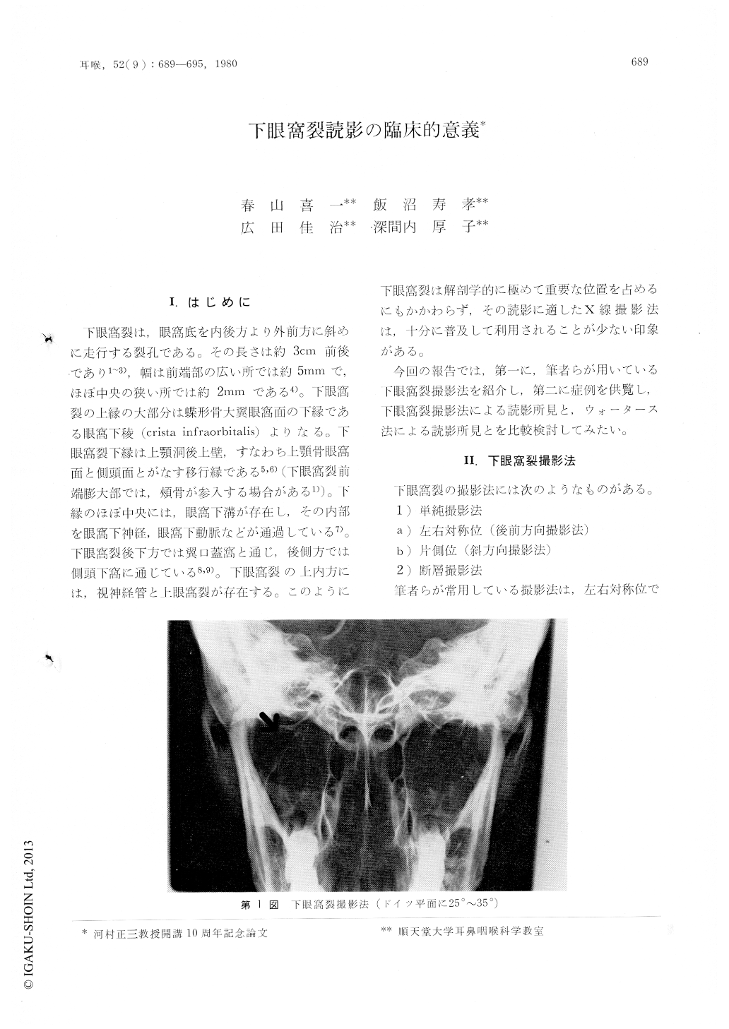
今回の報告では,第一に,筆者らが用いている下眼窩裂撮影法を紹介し,第二に症例を供覧し,下眼窩裂撮影法による読影所見と,ウォータース法による読影所見とを比較検討してみたい。
The inferior orbital fissure (IOF) is one of the important struttures in the surgical anatomy of the facial bone, and the radiological evaluation of IOF has not been well documented. Our investigation revealed that a PA view with the central ray of 25 to 35 degrees caudal to the Frankfort plane is the most appropriate view for the symmetrical evaluation of the IOF. The radiological evaluation of IOF is indicated for lesions involving the posterior aspect of the orbital floor or the posterior superior angle of the maxillary sinus. Important lesions are neoplasms and cystic lesions of the maxillary sinus.

Copyright © 1980, Igaku-Shoin Ltd. All rights reserved.


