Japanese
English
- 有料閲覧
- Abstract 文献概要
- 1ページ目 Look Inside
I.はじめに
頸部側面軟部撮影は喉頭,下咽頭の異常,甲状腺疾患その他の頸部の疾患に際してもつとも基本となる撮影で日常行なわれている。X線解剖的には気道の輪廓,咽頭後部,気管後部の軟部陰影,prevertebal fat stripe,頸部骨格ならびに骨化した喉頭あるいは気道軟骨が重要である。骨化喉頭軟骨に関しては形態観察を年齢別,性別に行ないその臨床的意義についてすでに発表した1)。
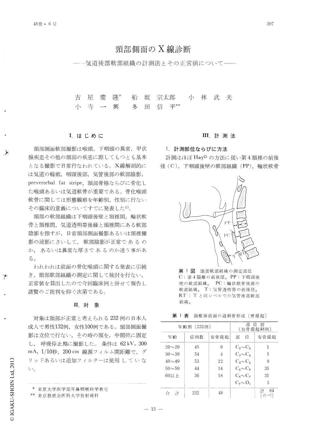
頸部の軟部組織は下咽頭後壁と頸椎間,輪状軟骨と頸椎間,気道透明帯後縁と頸椎間にある軟部陰影を指すが,日常頸部側面撮影あるいは頸椎撮影の読影にさいして,軟部陰影が正常であるのか,あるいは異常な厚さであるのか迷う事がある。
The X-ray films of the neck lateral view werestudied on normal human adults of 132 male and 100 females for interperetation of the soft tissue, e. g. retropharyngeal, postcricoid and posttracheal tissues, and the following conclusions were obtained.
1. The normal value of thickness of the retropharyngeal tissues at the hypopharynx was within 6 mm.
2. The normal thickness of the soft tissue shadow at postcricoid part was within 9 mm.
3. The normal width between the posterior wall of the trachea and anterior edge of the vertebra was within 15 mm. in male and 13 mm. in female.
4. The ratio between the width stated above and of inner diameter of the trachea was 1 or smaller than 1.
The patients with a retropharyngeal abscess, an esophageal tumor, a thyroid tumor and a tuberculosis of the cervical vertebra were examined and all of them showed abnormal values in these mesures.

Copyright © 1976, Igaku-Shoin Ltd. All rights reserved.


