Japanese
English
- 有料閲覧
- Abstract 文献概要
- 1ページ目 Look Inside
I.緒言
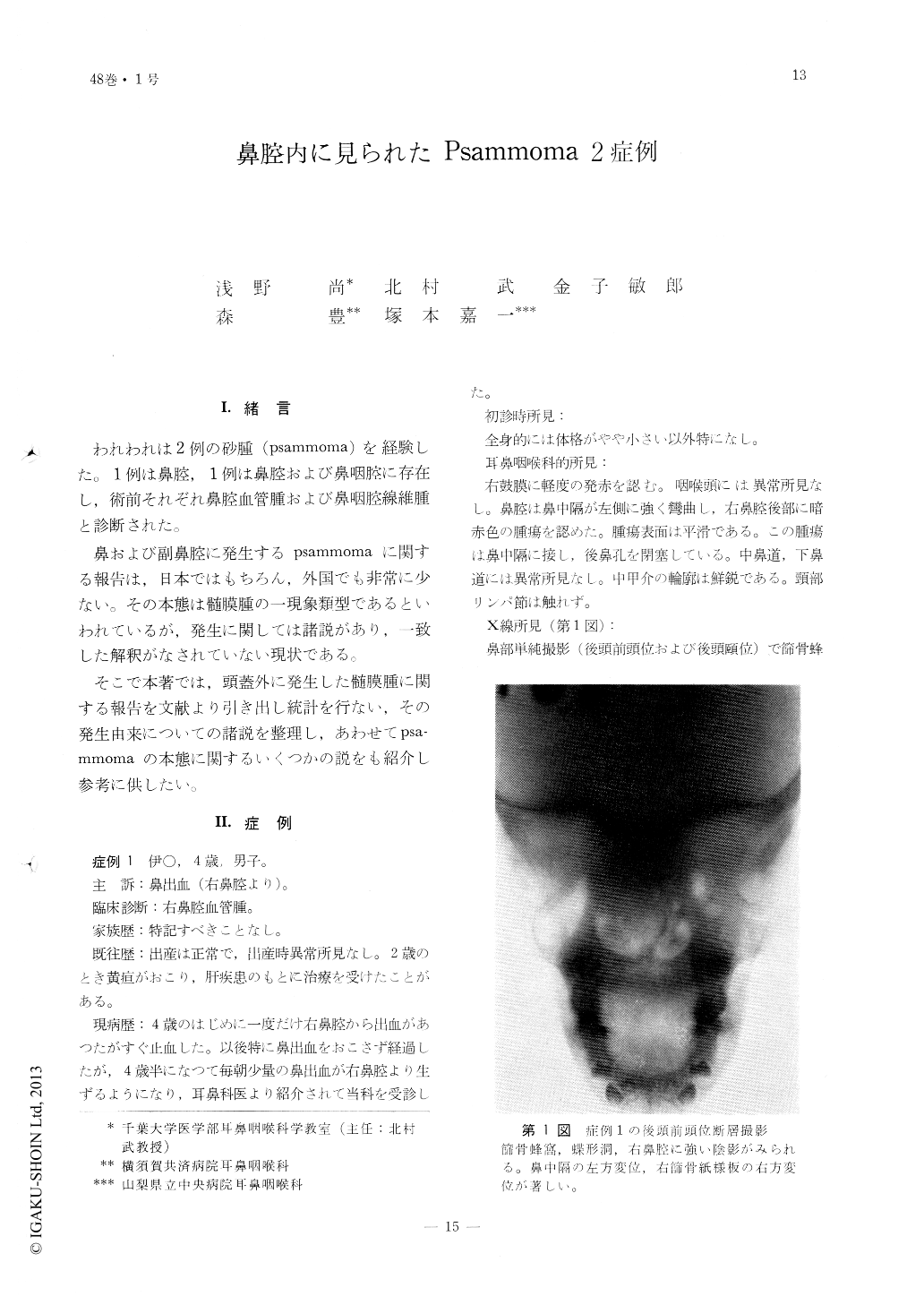
われわれは2例の砂腫(psammoma)を経験した。1例は鼻腔,1例は鼻腔および鼻咽腔に存在し,術前それぞれ鼻腔血管腫および鼻咽腔線維腫と診断された。
鼻および副鼻腔に発生するpsammomaに関する報告は,日本ではもちろん,外国でも非常に少ない。その本態は髄膜腫の一現象類型であるといわれているが,発生に関しては諸説があり,一致した解釈がなされていない現状である。
Two cases of psammoma, one of the nasal cavity, and the other of the paranasal sinus, are reported.
Case 1. A boy 4 year old complained of recurrent epistaxis. Nasal examination revealed a large tumor, darkred in color, filling the right nasal cavity and extending to the choana. A clinical diagnosis of hemangioma was made. The operation was perfomed by arched skin incision in the eyebrow; the sphenoid was found to be filled with the tumor mass which was fragile to touch and because of the profuse bleeding of this mass its removal was quite difficult. Microscopically, a large number of "psammoma bodies" were found in the tissues. These bodies were eosinophilic and the fibroblast-like cells with spindly nuclei were also increased in number.
Case 2. A man, aged 24, complained of obstruction of the left nasal cavity, hyposmia and epiphora for the past 7 years. Examination revealedthe nasal cavity and the corresponding choana filled with fibrous tumor mass and the left eye-brow pushed upwards about 5 mm.
A preoperative diagnosis of nasopharyngeal fibroma was made.
The operation was performed by Matis' skin incision. A very large tumor filling completely the nasal cavity and encroaching upon the nasal septum as well as the lateral wall of nasal cavity and corresponding contents of orbita that caused dislocation of its contents, were found. The tumor was resected together with the lateral wall of the nasal cavity as well as the middle and inferior conchae.
Microscopically, eosinophilic psammoma bodies were numerously recognized in the tissue.

Copyright © 1976, Igaku-Shoin Ltd. All rights reserved.


