Japanese
English
- 有料閲覧
- Abstract 文献概要
- 1ページ目 Look Inside
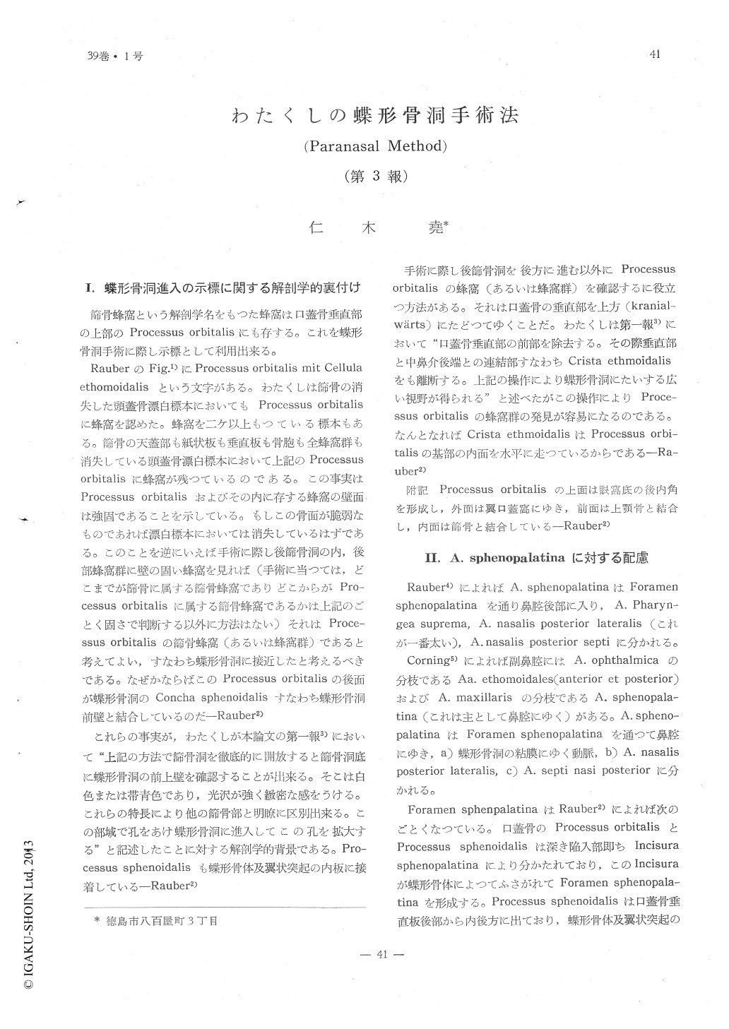
I.蝶形骨洞進入の示標に関する解剖学的裏付け
筋骨蜂窩という解剖学名をもつた蜂窩は口蓋骨垂直部の上部のProcessus orbitalisにも存する。これを蝶形骨洞手術に際し示標として利用出来る。
RauberのFig. 1)にProcessus orbitalis mit Cellula ethomoidalisという文字がある。わたくしは節骨の消失した頭蓋骨漂白標本においてもProcessus orbitalisに蜂窩を認めた。蜂窩を二ケ以上もつている標本もある。篩骨の天蓋部も紙状板も垂直板も骨胞も全蜂窩群も消失している頭蓋骨漂白標本において上記のProcessusorbitalisに蜂窩が残つているのである。この事実はProcessus orbitalisおよびその内に存する蜂窩の壁面は強固であることを示している。もしこの骨面が脆弱なものであれば漂白標本においては消失しているはずである。このことを逆にいえば手術に際し後鯖骨洞の内,後部蜂窩群に壁の固い蜂窩を見れば(手術に当つては,どこまでが篩骨に属する篩骨蜂窩でありどこからがProcessus orbitalisに属する篩骨蜂窩であるかは上記のごとく固さで判断する以外に方法はない)それはProcessus orbitalisの篩骨蜂窩(あるいは蜂窩群)であると考えてよい,すなわち蝶形骨洞に接近したと考えるべきである。なぜかならばこのProcessus orbitalisの後面が蝶形骨洞のConcha sphenoidalisすなわち蝶形骨洞前壁と結合しているのだ―Rauber2)
I have reported, elsewhere, that, after removal of the ethmoidal cells, the approach for opening the sphenoid sinus should be selected at a point close to the base of the former structure which is shown by an area different in color and luster from surrounding parts. Anatomically, this area is composed of cells in the processus orbitalis of the palatine bone. I try to avoid incurring injury to the sphenopalatine artery by closely adhering to the perpendicular portion of the palatine bone circumbenting touching the soft tissue adjacent to the sphenopalatine foramen. Another useful guide in locating the locus of entry is to find the posterior border of the perpendicular plate of the ethmoid because it is in line as a direct opposition to the crista sphenoidalis.

Copyright © 1967, Igaku-Shoin Ltd. All rights reserved.


