Japanese
English
--------------------
慢性中耳炎肉芽の電子顕微鏡的研究
A STUDY ON THE GRANULATION TISSUES IN CHRONIC OTITIS MEDIA THROUGH AN ELECTRONIC MICROSCOPE
中井 義明
1
Yoshiaki Nakai
1
1大阪市立大学耳鼻咽喉科学教室
pp.199-207
発行日 1965年3月20日
Published Date 1965/3/20
DOI https://doi.org/10.11477/mf.1492203397
- 有料閲覧
- Abstract 文献概要
- 1ページ目 Look Inside
Ⅰ.微細構造について
1.緒言
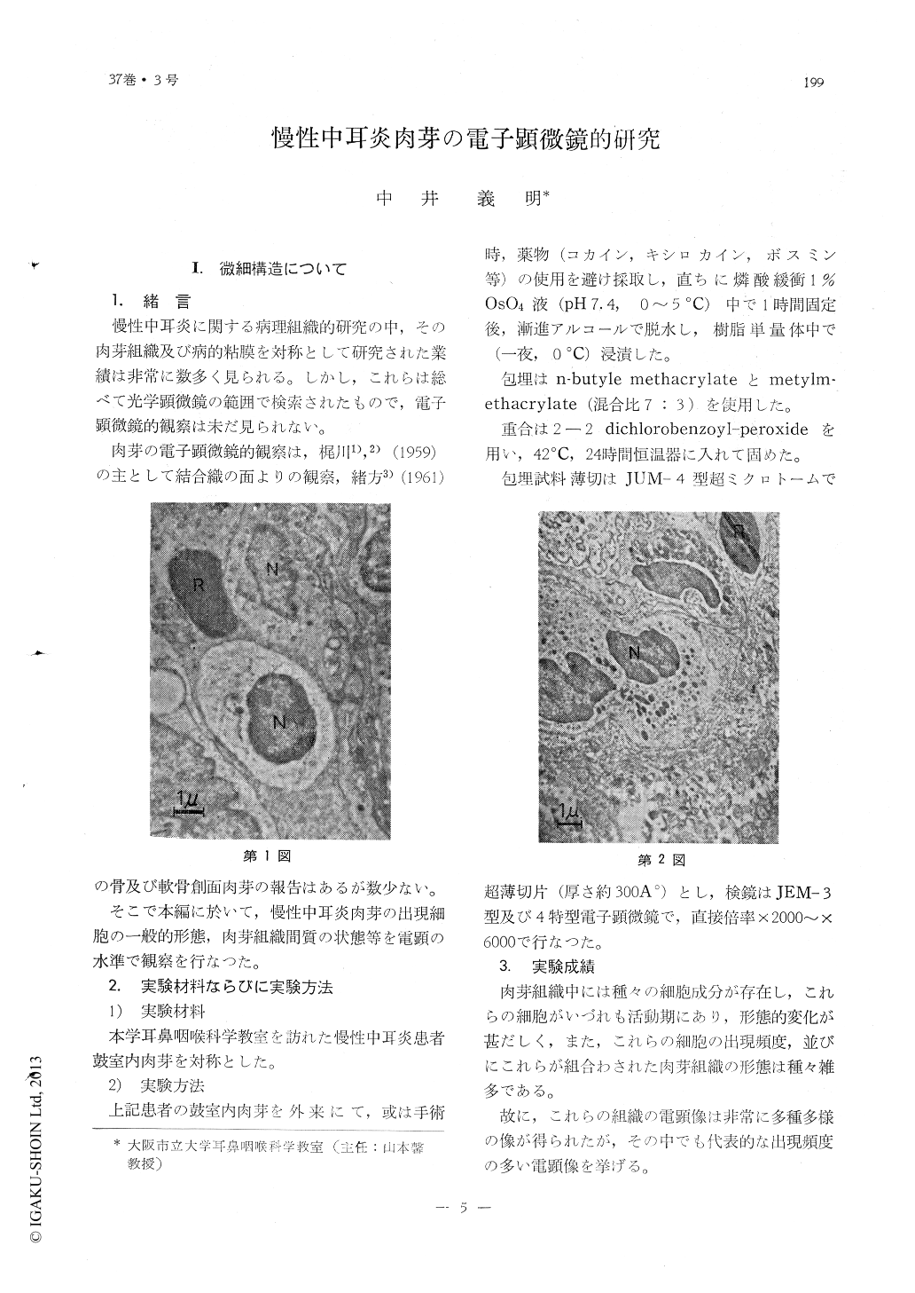
慢性中耳炎に関する病理組織的研究の中,その肉芽組織及び病的粘膜を対称として研究された業績は非常に数多く見られる。しかし,これらは総べて光学顕微鏡の範囲で検索されたもので,電子顕微鏡的観察は未だ見られない。
肉芽の電子顕微鏡的観察は,梶川1),2)(1959)の主として結合織の面よりの観察,緒方3)(1961)の骨及び軟骨創面肉芽の報告はあるが数少ない。
The cell structures of granulation tissues found in the otitis media were observed thr-ough electronic microscopy which revealed various stages of inflammatory process rang-ing from that of the early to the cured.
The mode in which the drugs or other inj-ected material permeate the individual cells of the granulation tissue were studied by means of using a tracer in 5% dextran iron solution and the surface activating solution (alevaire).

Copyright © 1965, Igaku-Shoin Ltd. All rights reserved.


