Japanese
English
症例
Diploic epidermoid cystの1例:画像診断を中心に
Diploic Epidermoid Cyst : Case Report
中塚 博貴
1
,
松原 一郎
1
Hiroki NAKATSUKA
1
,
Ichirou MATSUBARA
1
1済生会西条病院脳神経外科
1Department of Neurological Surgery, Saiseikai Saijo Hospital
キーワード:
epidermoid cyst
,
diploic
,
skull tumor
,
magnetic resonance imaging
,
diffusion-weighted image
Keyword:
epidermoid cyst
,
diploic
,
skull tumor
,
magnetic resonance imaging
,
diffusion-weighted image
pp.57-61
発行日 2003年1月10日
Published Date 2003/1/10
DOI https://doi.org/10.11477/mf.1436902332
- 有料閲覧
- Abstract 文献概要
- 1ページ目 Look Inside
Ⅰ.はじめに
類表皮嚢胞は全脳腫瘍の0.5〜1.5%を占める腫瘍で9),そのうち頭蓋骨の板間層に発生するものは16.2%と比較的稀であるが8),頭蓋骨腫瘍をみた際に鑑別に考慮しておくべき疾患の1つである.今回われわれは,板間層類表皮嚢胞の1例を経験したので,その画像的特徴などに関する文献的考察を加えて報告する.
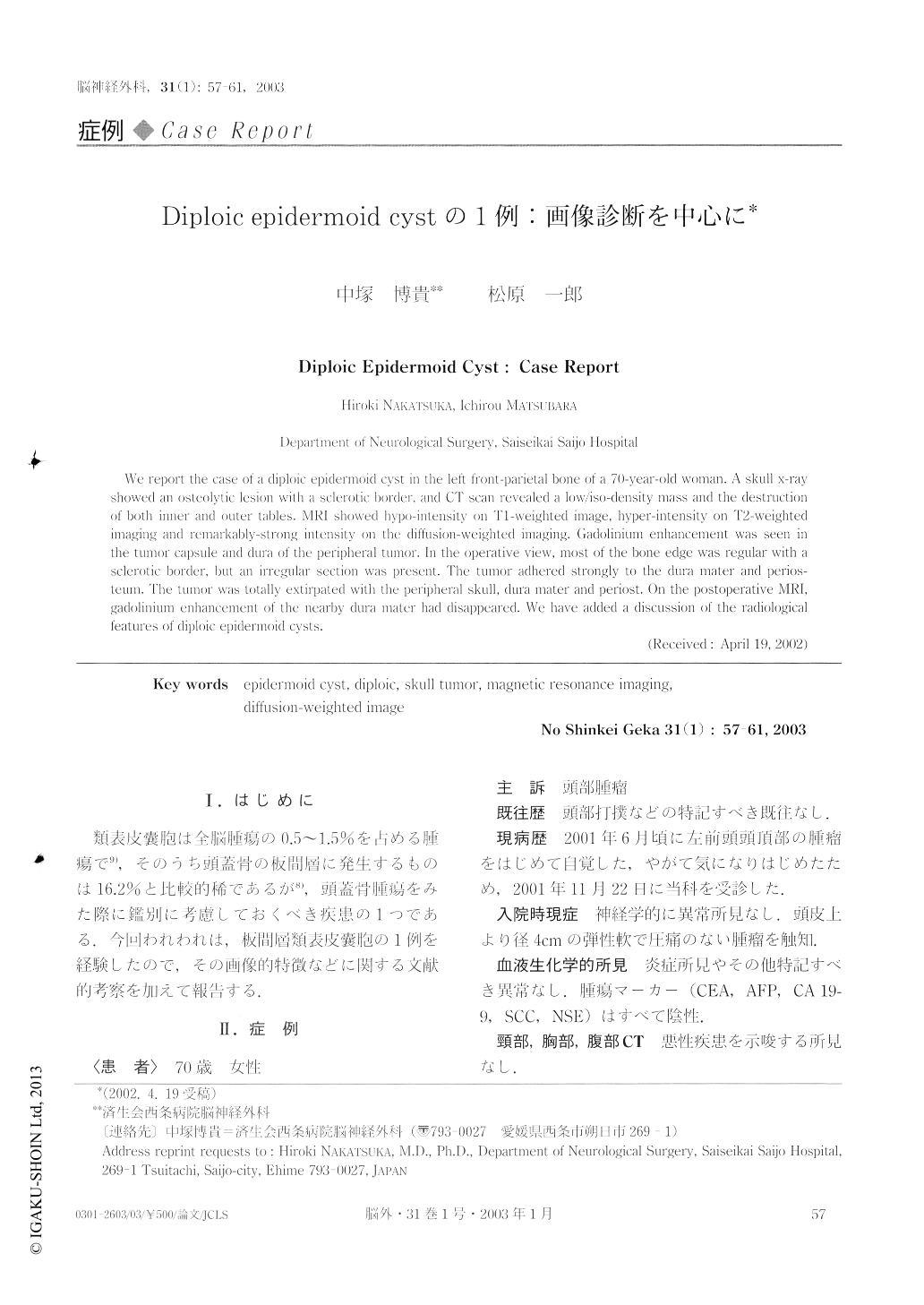
We report the case of a diploic epidermoid cyst in the left front-parietal bone of a 70-year-old woman. A skull x-ray showed an osteolytic lesion with a sclerotic border, and CT scan revealed a low/iso-density mass and the destruction of both inner and outer tables. MRI showed hypo-intensity on TI-weighted image, hyper-intensity on T2-weighted imaging and remarkably-strong intensity on the diffusion-weighted imaging. Gadolinium enhancement was seen in the tumor capsule and dura of the peripheral tumor.

Copyright © 2003, Igaku-Shoin Ltd. All rights reserved.


