Japanese
English
- 有料閲覧
- Abstract 文献概要
- 1ページ目 Look Inside
1.はじめに
中心溝近傍に存在する腫瘍性病変に関して,術前にその解剖学的局在を明確にしておくことは,安全かつ的確な手術において有用であり,func-tional magnetic resonance imaging(fMRI)を用いた非侵襲的画像検査が術前の機能マッピングとして試みられている1,7).一方,transcranial magne-tic stimulation(TCS)はベットサイドにおいて簡便かつ無侵襲に四肢の筋運動誘発が可能であり,術前の運動中枢マッピングへの応用も検討されている9).前者は神経活動負荷にともなう過剰なオキシヘモグロビンの増加,即ち血流の増加を捉えることで,機能局在を間接的に把握する方法である11).後者では磁気刺激によって脳内に生じた誘導電流が神経細胞を刺激し,その支配の筋運動が誘発されることで,直接的に機能局在の決定が可能である3).しかし刺激用コイルと頭皮面接線との角度の影響など刺激手技によってマーキングに誤差を生じる危険性を有している.今回はfMRIおよびTCSによって得られた術前運動野同定所見を比較検討し,一部症例では術中所見とも対比しfMRIおよびTCSを用いた術前の運動野マッピングについて,その意義および問題点を報告した.
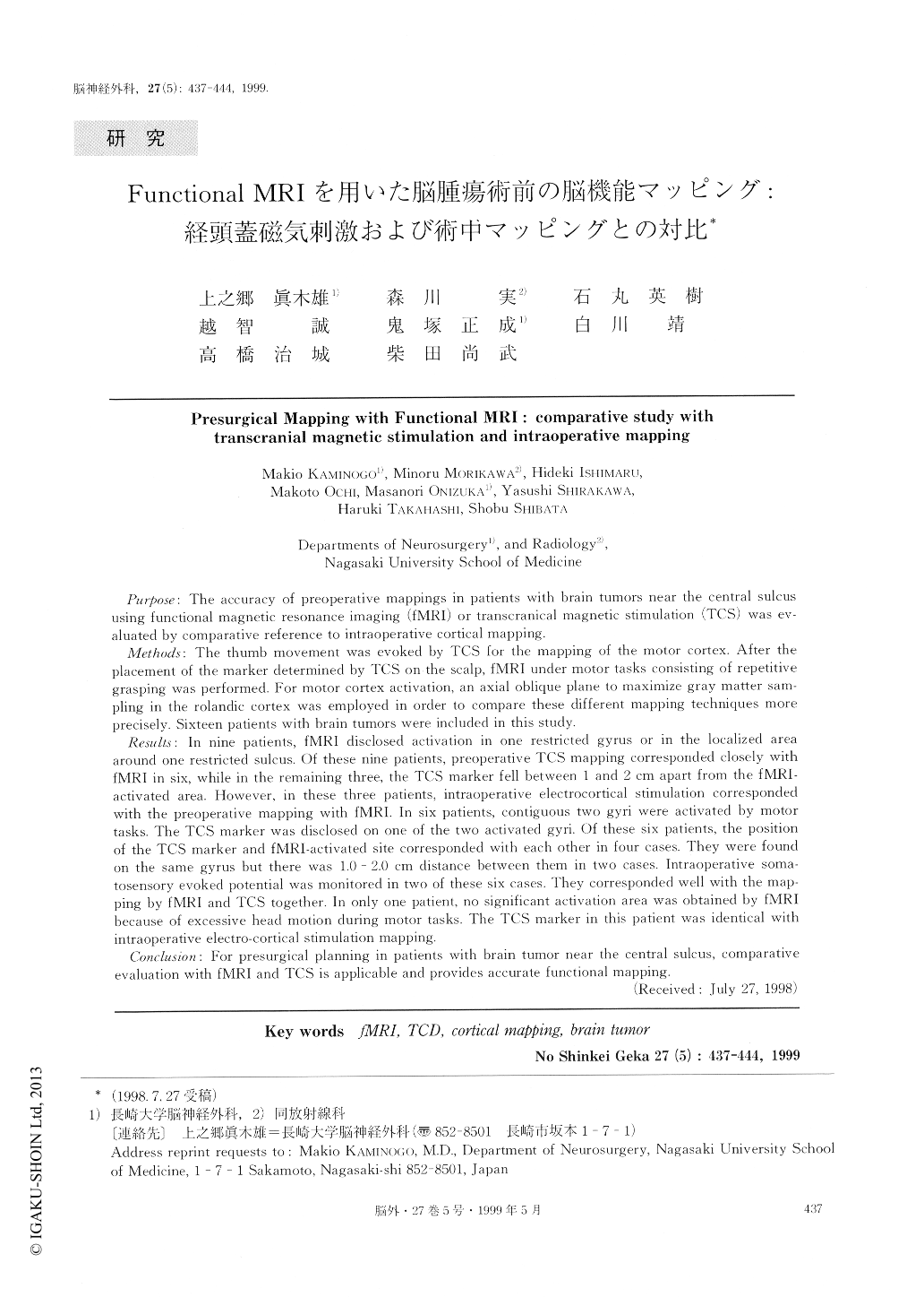
Purpose: The accuracy of preoperative mappings in patients with brain tumors near the central sulcus using functional magnetic resonance imaging (fMRI) or transcranical magnetic stimulation (TCS) was ev-aluated by comparative reference to intraoperative cortical mapping.
Methods: The thumb movement was evoked by TCS for the mapping of the motor cortex. After the placement of the marker determined by TCS on the scalp, fMRI under motor tasks consisting of repetitive grasping was performed. For motor cortex activation, an axial oblique plane to maximize gray matter sam-pling in the rolandic cortex was employed in order to compare these different mapping techniques more precisely. Sixteen patients with brain tumors were included in this study.
Results: In nine patients, fMRI disclosed activation in one restricted gyrus or in the localized area around one restricted sulcus. Of these nine patients, preoperative TCS mapping corresponded closely with fMRI in six, while in the remaining three, the TCS marker fell between I and 2 cm apart from the fMRI-activated area. However, in these three patients, intraoperative electrocortical stimulation corresponded with the preoperative mapping with fMRI. In six patients, contiguous two gyri were activated by motor tasks. The TCS marker was disclosed on one of the two activated gyri. Of these six patients, the position of the TCS marker and fMRI-activated site corresponded with each other in four cases. They were found on the same gyrus but there was 1.0 - 2.0 cm distance between them in two cases. Intraoperative soma-tosensory evoked potential was monitored in two of these six cases. They corresponded well with the map-ping by fMRI and TCS together. In only one patient, no significant activation area was obtained by fMRI because of excessive head motion during motor tasks. The TCS marker in this patient was identical with intraoperative electro-cortical stimulation mapping.
Conclusion: For presurgical planning in patients with brain tumor near the central sulcus, comparative evaluation with fMRI and TCS is applicable and provides accurate functional mapping.

Copyright © 1999, Igaku-Shoin Ltd. All rights reserved.


