Japanese
English
- 有料閲覧
- Abstract 文献概要
- 1ページ目 Look Inside
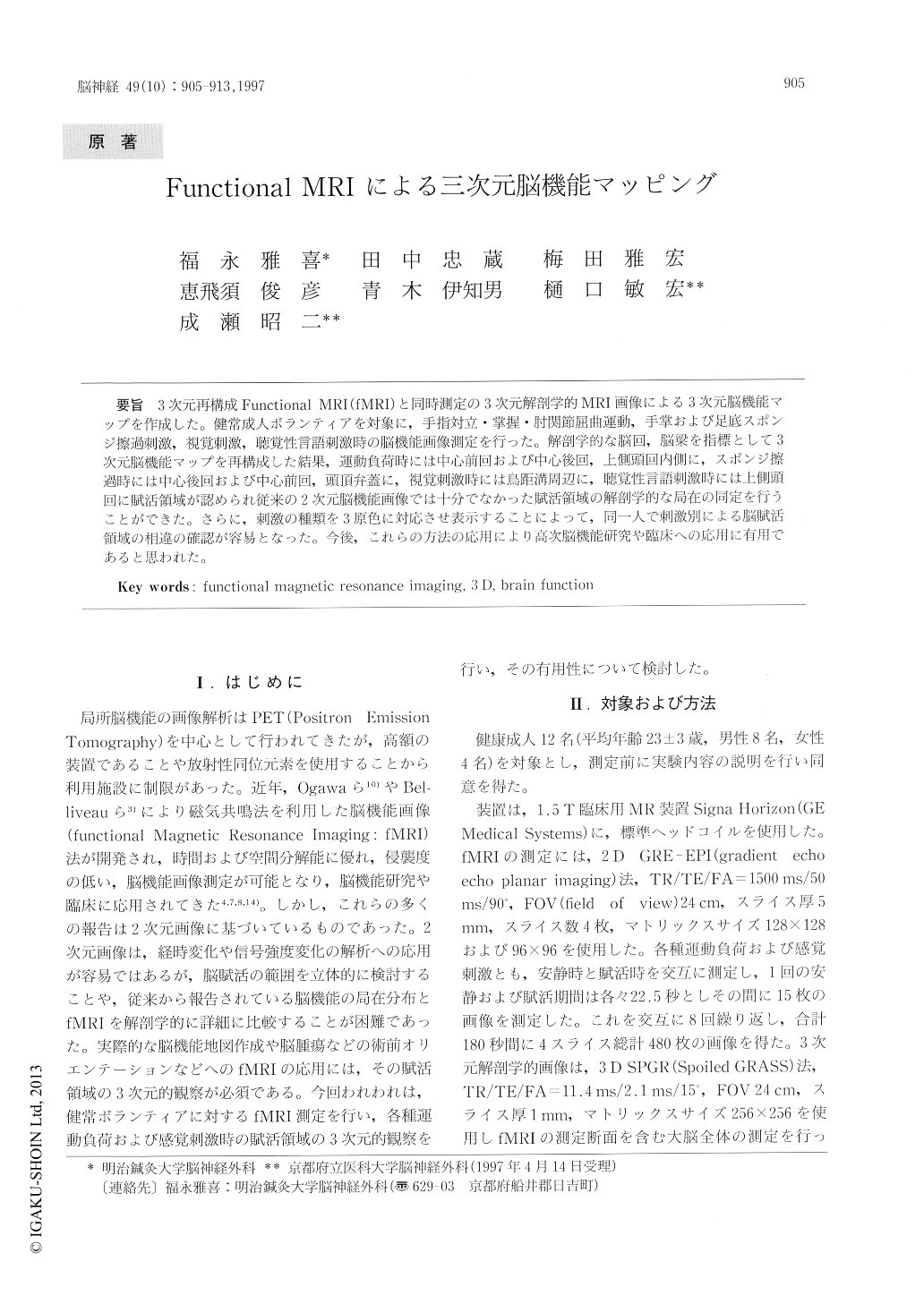
3次元再構成Functional MRI(fMRI)と同時測定の3次元解剖学的MRI画像による3次元脳機能マップを作成した。健常成人ボランティアを対象に,手指対立・掌握・肘関節屈曲運動,手掌および足底スポンジ擦過刺激,視覚刺激,聴覚性言語刺激時の脳機能画像測定を行った。解剖学的な脳回,脳梁を指標として3次元脳機能マップを再構成した結果,運動負荷時には中心前回および中心後回,上側頭回内側に,スポンジ擦過時には中心後回および中心前回,頭頂弁蓋に,視覚刺激時には鳥距溝周辺に,聴覚性言語刺激時には上側頭回に賦活領域が認められ従来の2次元脳機能画像では十分でなかった賦活領域の解剖学的な局在の同定を行うことができた。さらに,刺激の種類を3原色に対応させ表示することによって,同一人で刺激別による脳賦活領域の相違の確認が容易となった。今後,これらの方法の応用により高次脳機能研究や臨床への応用に有用であると思われた。
Functional mapping of the activated brain, the location and extent of the activated area were determined, during motor tasks and sensory stimu-lation using fMRI superimposed on 3 D anatomical MRI.
Twelve volunteers were studied. The fMR images were acquired using a 2 D gradient echo echo planarimaging sequence. The 3 D anatomical MR images of the whole brain were acquired using a conven-tional 3 D gradient echo sequence. Motor tasks were sequential opposition of fingers, clenching a hand and elbow flexion. Somatosensory stimulation were administered by scrubbing the palm and sole with a washing sponge.

Copyright © 1997, Igaku-Shoin Ltd. All rights reserved.


