Japanese
English
- 有料閲覧
- Abstract 文献概要
- 1ページ目 Look Inside
I.はじめに
Malignant fibrous histiocytoma(MFH)は,軟部組織のsarcomaでは最も多く,10.5-34%を占め,四肢,後腹膜に好発する4,6,29).一方,頭蓋内原発のMFHは,1976年にGonzalez-Vitaleらによって初めて報告されたが8),いまだ文献上散見するのみであり,極めて稀と思われる.今回,我々は,興味ある発育形態を示した頭蓋内原発MFHの1例を経験したので,若干の文献的考察を加えて報告する.
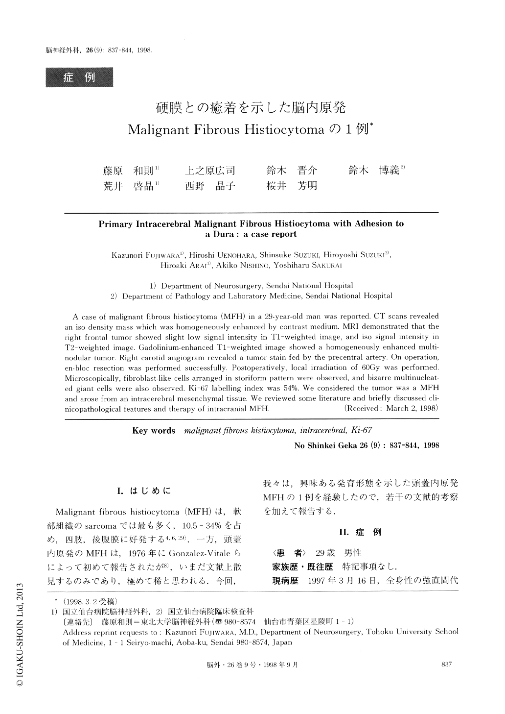
A case of malignant fibrous histiocytoma (MFH) in a 29-year-old man was reported. CT scans revealedan iso density mass which was homogeneously enhanced by contrast medium. MRI demonstrated that theright frontal tumor showed slight low signal intensity in T1-weighted image, and iso signal intensity inT2-weighted image. Gadolinium-enhanced T1-weighted image showed a homogeneously enhanced multi-nodular tumor. Right carotid angiogram revealed a tumor stain fed by the precentral artery. On operation,en-bloc resection was performed successfully. Postoperatively, local irradiation of 60Gy was performed.Microscopically, fibroblast-like cells arranged in storiform pattern were observed, and bizarre multinucleat-ed giant cells were also observed. Ki-67 labelling index was 54%. We considered the tumor was a MFHand arose from an intracerebral mesenchymal tissue. We reviewed some literature and briefly discussed cli-nicopathological features and therapy of intracranial MFH.

Copyright © 1998, Igaku-Shoin Ltd. All rights reserved.


