Japanese
English
- 有料閲覧
- Abstract 文献概要
- 1ページ目 Look Inside
I.はじめに
脳腫瘍に対する放射線治療後に脳血管の狭窄もしくは閉塞を来たすことがあり1,3,8,10),もやもや病と類似の血管写所見を呈する類もやもや12)も報告されている.今回われわれは放射線治療11年後に再発した頭蓋咽頭腫で類もやもやを合併していた症例に対し,開頭手術を行わずに経蝶形骨洞手術とガンマナイフによる治療を行い良好な結果を得た.その臨床経過を報告するとともに,放射線治療後の類もやもやを伴った再発頭蓋咽頭腫に対する治療法の選択について考察する.
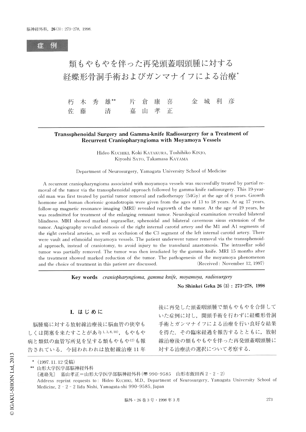
A recurrent craniopharyngioma associated with moyamoya vessels was successfully treated by partial re-moval of the tumor via the transsphenoidal approach followed by gamma-knife radiosurgery. This 19-year-old man was first treated by partial tumor removal and radiotherapy (54Gy) at the age of 6 years. Growthhormone and human chorionic gonadotropin were given from the ages of 13 to 18 years. At ag 17 years,follow-up magnetic resonance imaging (MRI) revealed regrowth of the tumor. At the age of 19 years, hewas readmitted for treatment of the enlarging remnant tumor. Neurological examination revealed bilateralblindness. MRI showed marked suprasellar, sphenoidal and bilateral cavernous sinus extension of thetumor. Angiography revealed stenosis of the right internal carotid artery and the M1 and A1 segments ofthe right cerebral arteries, as well as occlusion of the C3 segment of the left internal carotid artery. Therewere vault and ethmoidal moyamoya vessels. The patient underwent tumor removal via the transsphenoid-al approach, instead of craniotomy, to avoid injury to the transdural anastomosis. The intrasellar solidtumor was partially removed. The tumor was then irradiated by the gamma knife. MRI 15 months afterthe treatment showed marked reduction of the tumor. The pathogenesis of the moyamoya pllenomenonand the choice of treatment in this patient are discussed.

Copyright © 1998, Igaku-Shoin Ltd. All rights reserved.


