Japanese
English
- 有料閲覧
- Abstract 文献概要
- 1ページ目 Look Inside
I.はじめに
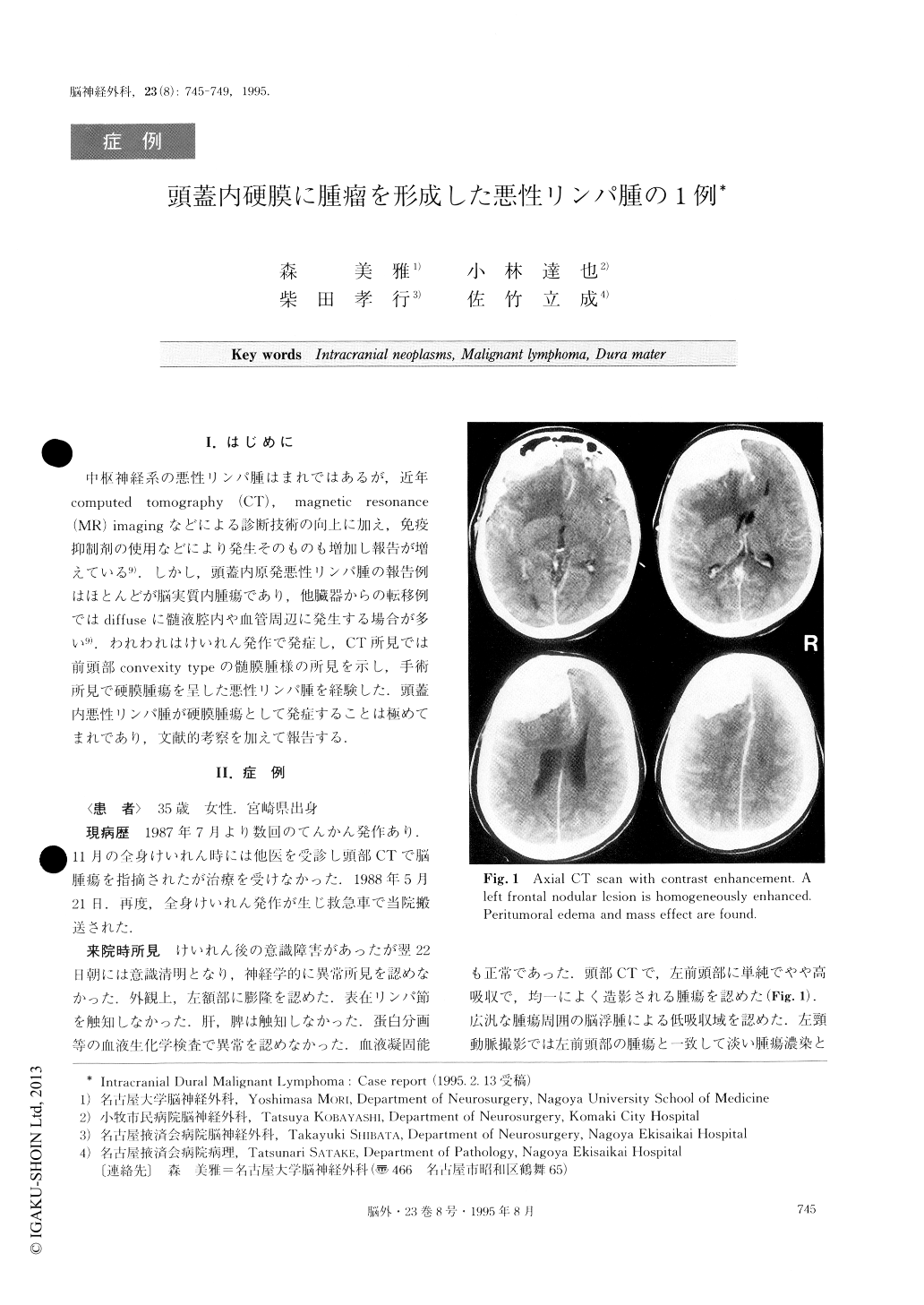
中枢神経系の悪性リンパ腫はまれではあるが,近年computed tomography(CT),magnetic resonance(MR)imagingなどによる診断技術の向上に加え,免疫抑制剤の使用などにより発生そのものも増加し報告が増えている9).しかし,頭蓋内原発悪性リンパ腫の報告例はほとんどが脳実質内腫瘍であり,他臓器からの転移例ではdiffuseに髄液腔内や血管周辺に発生する場合が多い9).われわれはけいれん発作で発症し,CT所見では前頭部convexity typeの髄膜腫様の所見を示し,手術所見で硬膜腫瘍を呈した悪性リンパ腫を経験した.頭蓋内悪性リンパ腫が硬膜腫瘍として発症することは極めてまれであり,文献的考察を加えて報告する.
A case of intracranial chiral malignant lymphoma was presented. A 35-year-old woman was admitted with generalized seizure. The neurological findings and laboratory data were normal. Computed tomography (CT) showed a nodular lesion in the left frontal con-vexity with marked enhancement by contrast medium.Angiography showed a slight tumor stain and the feed-ing artery from the ethmoid artery. On surgery, a soft pinkish tumor was found to be located in the dura and subdural space. It was resected subtotally. The histolo-gical diagnosis was malignant lymphoma of B cell ori-gin. Postoperative Ga scintigraphy and abdominal CT disclosed no abnormalities in other organs, but bone marrow examination revealed lymphoma cells, which suggested that the patient might have systemic lympho-ma. The patient and her family refused additional treat-ment. The patient is alive and well now over six years after the craniotomy. Primary central nervous system (CNS) lymphomas usually arise in the cerebral paren-chyma, while secondary CNS lymphomas commonly in-volve leptomeninges and perivascular spaces. Mass le-sions arising in the dura are very rare.

Copyright © 1995, Igaku-Shoin Ltd. All rights reserved.


