Japanese
English
- 有料閲覧
- Abstract 文献概要
- 1ページ目 Look Inside
I.はじめに
頭蓋内原発性悪性リンパ腫は比較的稀な疾患とされてきたが,画像診断の発達や移植やそれに伴う免疫抑制剤の使用,および免疫学的知見の進歩などにより症例の報告数が増加し注目されてきている.また,それらの報告のほとんどがB細胞である3,7,13).しかし,中枢神経系以外の悪性リンパ腫では,非典型的な細胞性格を呈する概念も確立されつつある.その中で,最近われわれはT-cell-rich B-cell lymphomaと考えられた頭蓋内原発性悪性リンパ腫の1剖検例を経験したので,若干の文献的考察を加えて報告する.
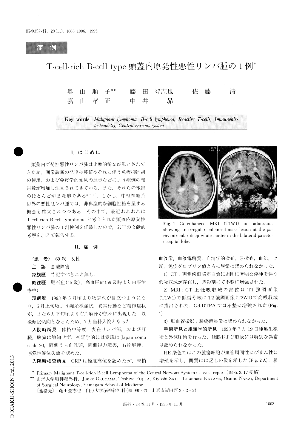
An autopsy case of primary intracranial T-cell-rich B-cell lymphoma in a 69-year-old female is presented. The patient was admitted with a diagnosis of a brain tumor in July 1993 and a month long history of mental de-terioration, motor weakness of the right arm and leg, and a tendency toward somnolence. Neurological ex-amination revealed disturbance of consciousness, right hemiparesis, and papilloedema. However, her general physical examination was unremarkable. A CT scan and MR imaging revealed an irregular enhanced mass lesion at the paraventricular deep white matter in the bilateral parieto-occipital lobe. The patient was treated with surgical biopsy of the tumor followed by com-bined radiotherapy (a total of 50 Gy) and chemother-apy. Following repetitive episodes of remission and ex-acerbation, the patient expired about seven months af-ter the onset of symptoms. Histopathological diagnosis of the tumor was malignant lymphoma (diffuse medium-sized cell type). In the immunohistochemical study, most of the lymphoma cells had T-cell markers, such as UCHL1. Some of the lymphoma cells were L26-positive. Neither glial fibrillary acidic protein nor neuron specific enolase were reactive with the lympho-ma cells. At post-mortem examination, the specimens disclosed diffuse infiltration of medium-sized lymphoma cells. By contrast, most of the lymphoma cells were shown to be positive by the analysis of L26. None of the lymphoma cells exhibited the presence of UCHL1. These immunohistochemical evaluations conform to the criteria of T-cell-rich B-cell lymphoma.

Copyright © 1995, Igaku-Shoin Ltd. All rights reserved.


