Japanese
English
- 有料閲覧
- Abstract 文献概要
- 1ページ目 Look Inside
I.はじめに
類上皮腫は頭蓋内腫瘍の約1%21)を占めるが,まれに悪性変化を伴うものがあり悪性類上皮腫もしくは類上皮腫癌と呼ばれ,予後は極めて不良である.われわれは摘出後,放射線治療による13カ月の症状軽快の後,再び急速に増大する小脳橋角部悪性類上皮腫に対してradiosurgeryを行った.その効果を含め悪性類上皮腫の治療に関して文献的考察を行う.
A case of epidermoid carcinoma in the cerebello-pontine (CP) angle is presented.
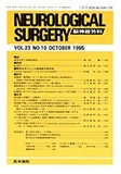
A 42-year-old male was admitted with a complaint of experiencing double vision for four months in January, 1992. During neurological examination, right abducens palsy, right facial dysesthesia, and atrophy of the right temporal muscle were noted. Magnetic resonance (MR) imaging revealed a mass of low intensity in the rightCP angle, which was prominently enhanced with gado-linium. Malignancy was suspected because the tumor on MR enlarged rapidly in a month, so the first surgical resection was performed. Suboccipital exploration of the right CP angle was performed in February. At first, a fragile, pearly part of the mass typical of epidermoid was exposed behind the seventh and eighth cranical nerve complex. Then, a grayish, fibrous part was ex-posed, which involved the fifth cranial nerve and was attached to the tentorium and the brainstem. Histologic-al diagnosis of the fragile part of the tumor revealed a typical epidermoid cyst and that of the fibrous part was squamous cell carcinoma. During postoperative ex-aminations on other parts of the body, such as endosco-pic studies of the trachea and the esophagus, no abnor-mality was shown. Therefore the tumor was diagnosed as a primary intracranial epidermoid carcinoma. Post-operatively, conventional fractionated external-beam focal irradiation was carried out, which caused regres-sion of the residual tumor for eleven months. Subse-quently, palsy of the right side of the tongue and pare-sis of the contralateral side of the extremities and face developed with increase of the right abducens palsy. MR imaging indicated regrowth of the tumor. The second operation via the subtemporal approach was un-successful, because the tumor was fibrous and firmly attached to the brainstem. Finaly, gamma radiosurgery was applied to the tumor at the end of April, where a marginal dose of 14 Gy was delivered. MR image taken three months after the radiosurgery showed the tumor unchanged in size, but with central loss of contrast en-hancement. Since four months after the radiosurgery, the tumor had shown rapid re-growth from the margin, with deterioration of the patient's neurological condi-tion. He died in April, 1994.
Copyright © 1995, Igaku-Shoin Ltd. All rights reserved.