Japanese
English
- 有料閲覧
- Abstract 文献概要
- 1ページ目 Look Inside
I.はじめに
転移性脳腫瘍は剖検では全悪性腫瘍の10-30%にみられ1,3,17,18),原発巣としては肺癌,乳癌,消化器系の癌が多い.—方転移性脳腫瘍の原発巣不明例は0.5-8%にみられ5,8,22,28),このうち剖検によっても原発巣が特定できない症例が15-27%ある5,6,8,15,21).このように原発巣不明の転移性脳腫瘍は臨床において時に遭遇する.われわれは,非定型的な特徴をもつ分化型腺癌で3回の頭蓋内腫瘍摘出術を受け,7年の間隔をあけた2度の全身検索でも原発巣が不明であった転移性脳腫瘍と考えられる長期生存例を経験したので,若干の文献的考察を加え報告する.
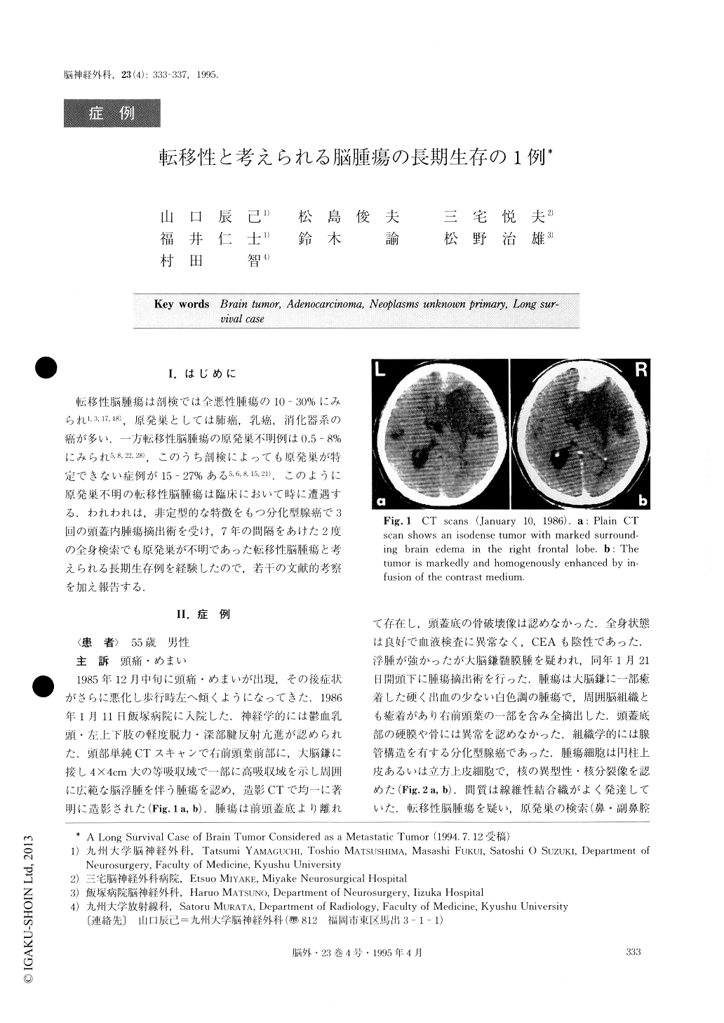
A case of long survival of brain tumor (well diffe-rentiated adenocarcinoma) was reported. A 55-year-old man was admitted in January, 1986, because of a one month history of progressive headache, dizziness and gait disturbance. CT scans revealed an enhancing tumor in contact with the falx in the right frontal lobe. The tumor was totally removed. The histopathological diagnosis was that of a well differentiated adenocarci-noma. The primary site of the adenocarcinoma was not detected. No chemotherapy or radiation therapy was given.
Four years and 7 months after surgery CT scans demonstrated a recurrent tumor as a bilaterally expand-ing falx meningioma. Nearly total removal of the tumor was again performed and diagnosed as adenocarcino-ma. Examinations to detect the primary site and other metastatic lesions were negative again. On May 1993, the patient died because of the intracranial dissemina-tion of tumor without extracranial lesions. The period from the first operation to his death was 7 years and 5 months.
This is a case of long survival of intracranial cancer, which was considered as a metastatic tumor, though the primary site and other metastatic lesions were not detected. The tumor in this case showed the atypical features of a metastatic adenocarcinoma. For example, the primary and recurrent tumors resembled a para-sagital or falx meningioma in shape and they grew slowly. Therefore, there is a possibility that the tumor was actually a primary adenocarcinoma, which might have arisen from the embryologically migrated cells of the mucous membrane or from ectopic epithelial cells.

Copyright © 1995, Igaku-Shoin Ltd. All rights reserved.


