Japanese
English
- 有料閲覧
- Abstract 文献概要
- 1ページ目 Look Inside
I.はじめに
最近,臨床で運動機能を評価するために経頭蓋大脳磁気刺激が盛んに使用されるようになってきた3,6,11,12,18-20,23).脳神経外科の領域では,中心溝付近の開頭手術を安全に施行するため,術前に頭皮上から運動領野あるいは中心溝を同定するために用いられている7,8,15).臨床的に問題となるのは,経頭蓋的磁気刺激の対象となる患者のほとんどが運動麻痺を伴っているため,正常人に比較して運動誘発電位(MEP)を得ることが困難であるという点である.特に上肢よりも下肢領域の運動誘発電位を得にくいため,下肢筋の運動領野を同定できないことが多く,麻痺患者では上肢から下肢までの運動領野全体をとらえにくい.
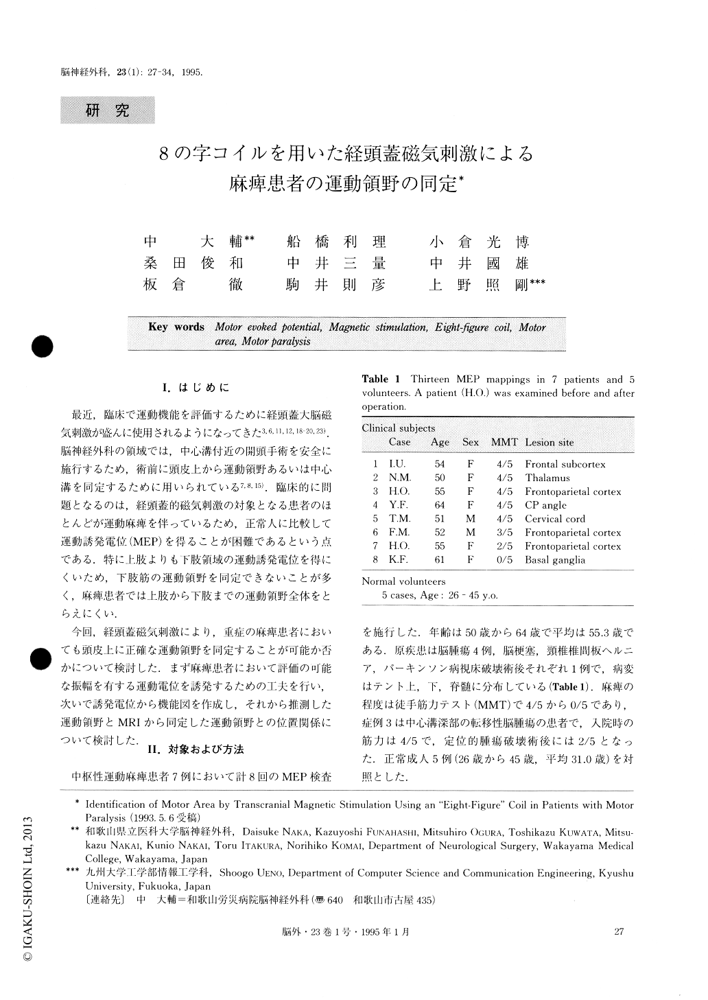
今回,経頭蓋磁気刺激により,重症の麻痺患者においても頭皮上に正確な運動領野を同定することが可能か否かについて検討した.まず麻痺患者において評価の可能な振幅を有する運動電位を誘発するための工夫を行い,次いで誘発電位から機能図を作成し,それから推測した運動領野とMRIから同定した運動領野との位置関係について検討した.
In patients with motor paralysis, we tried to indentify the functional motor area by transcranial magnetic sti-mulation using an “eight-figure” coil designed by Ueno.
Motor evoked potentials (MEPs) were recorded in 7 patients from 50 to 64 years old, and in 5 normal volun-teers 26 to 45 years old. They were stimulated at 49 points over an unilateral hand motor area, and at 21 points over a foot motor area, and surface MEPs were recorded on their contralateral thenar muscle and abductor hallucis brevis muscle. In normal volunteers, the optimal eddy current for stimulating the hand motor area was directed anteriorly parallel to the mid-line, and for stimulating the foot motor area, it was postero-laterally directed with an angle of 45 degrees towards the midline. MEPs could be induced at their muscle contractions during which their thumbs and middle fingers softly touched each other, and their hal-luxes slightly flexed. In five patients two kinds of am-plitude mappings reconstructed from MEPs were obtained at rest or at muscle contraction. A line con-necting these two peaks on an amplitude mapping was regarded as an “MEP-motor area”. A geographical dif-ference between the MEP-motor area and MRI-motor area (identified by an MRI surface image) was studied at muscle contraction and at rest.
In normal subjects the sites of the MEP-motor area and of the MRI-motor area coincided, whereas, in pa-tients with space-occupying lesions near the central sul-cus, the MEP-motor areas were located 1 to 2 cm pos-terior to MRI-motor areas. Motor area was detected in 2 MEP mappings recorded at rest and in 3 mappings at voluntary muscle contraction. Another 3 mappings failed to show any MEP peaks either at rest or at mus-cle contraction.
Two of 3 cases whose mappings did not show any MEPs even during the muscle contraction had muscle weakness (2/5 and 0/5 in MMT). In all but one with milder motor paresis (stronger than 3/5 in MMT). MEP mappings showed prominent peaks at hand and foot areas.
Transcranial magnetic stimulation for detecting the motor area is easy to perform and painless for the pa-tients. It can present the precise location of the motor area in normal subjects and is regarded to be a useful method to detect the motor area from over the scalp. But in patients with lesions near central sulcus, it should be kept in mind that the MEP-motor area is slightly different from the MRI-motor area, probably clue to the dislocation or distortion of the pyramidal ax-ons. The activation of muscle contraction by transcra-nial magnetic stimulation is thought to be a useful man-euver to evoke MEPs and to detect the cortical motor area from over the scalp in patients with motor para-lysis.
Copyright © 1995, Igaku-Shoin Ltd. All rights reserved.