Japanese
English
- 有料閲覧
- Abstract 文献概要
- 1ページ目 Look Inside
I.はじめに
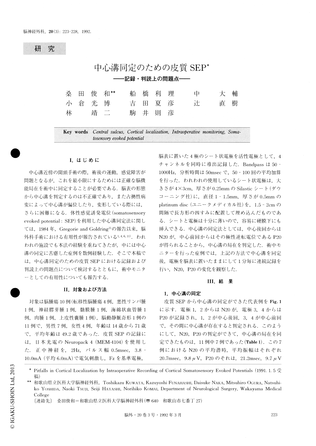
中心溝近傍の開頭手術の際,術後の運動,感覚障害が問題となるが,これを最小限にするためには正確な脳機能局在を術中に同定することが必要である.脳表の形態から中心溝を判定するのは不正確であり,また占拠性病変によって中心溝が偏位したり,変形している際には,さらに困難になる.体性感覚誘発電位(somatosenso)ryevoked potential:SEP)を利用した中心溝同定法に関しては,1984年,Gregorie and Goldring3)の報告以来,脳外科手術における有用性が報告されている1,4,6,11).われわれの施設でも本法の経験を重ねてきたが,中には中心溝の同定に苦慮した症例を数例経験した.そこで本稿では,中心溝同定のための皮質SEPにおける記録および判読上の問題点について検討するとともに,術中モニターとしての右用性についても報告する.
Cortical somatosensory evoked potential (SEP) re-cordings were made in 11 patients who had lesions lo-cated in or near the somatosensory or motor gyri to localize the central sulcus and sensorimotor cortex dur-ing neurosurgical operations. Cortical localization was successful in 7 of the 11 patients by recording phase re-versal waveforms of N20 and P20 at electrode sites in the hand area on opposite sides of the central sulcus. There were 4 cases in which the cortical localization failed. Locations of craniotomy were far distant from the central sulcus retrospectively in 2 of the 4 patients. Cortical SEPs couldn't be recorded despite probable ex-posure of the hand area and apparently adequate sti-mulation and recording conditions in 2 patients who had showed no or low amplitude scalp SEP preoper-atively. In one of these 2 patients only low amplitude negative waves were recorded at the cortex which was thought far field potentials originated from subcortical structures. In 2 patients cortical SEP was monitored during the removal of the tumors and was useful to estimate the effects of the operative procedures on the sensorimotor cortex.
It is concluded that the localization of cortical func-tions using cortical SEP is useful for reducing risk associated with intracranial surgery. However, we must be aware that there are some pitfalls in this method.

Copyright © 1992, Igaku-Shoin Ltd. All rights reserved.


