Japanese
English
- 有料閲覧
- Abstract 文献概要
- 1ページ目 Look Inside
I.はじめに
コンピュータを用いた立体画像化が進むなかで,脳血管の三次元表示が最近開発され,臨床的に応用されつつある1-5).しかし,使用されている機種は大型のコンピュータであり,設置するうえで経済的な問題があり,さらに三次元表示に要する時間がかかりすぎるという欠点があって臨床での利用価値が少ないのが現状である.これらの欠陥を補うべく,今回われわれは現在普及しているパーソナルコンピュータと市販のソフトプログラムを用いて,低価格でしかも迅速に脳血管の三次元画像を作成する方法を考案した.
Abstract
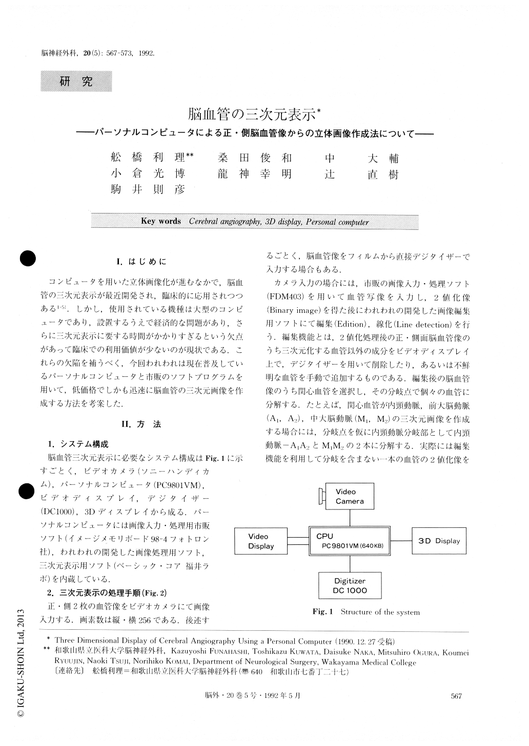
A new method for a 3-D reconstruction of angio-graphic cerebral vessels was developed. This system was constructed from inexpensive equipment such as a personal computer, a video camera, a video display, and a digitizer.
The input of the vessel images on anteroposterior and lateral angiograms was performed using a video camera with a personal computer and an image proces-sing program (FDM 98-4 ; Fotoron Co.). These proces-sed images were modified with our original editorial method. This can contribute a lot to the erasing of “non-vessel” images such as skull bone and other noise.It also adds concerning vessels which cannot be clearly taken from angiograms with the video camera. The most beneficial point of the editorial method was to re-duce the time consumed in the reconstruction. The spatial points of X-ray discharging boxes against the AP and lateral films were calculated from the length and width of the radiated images of metal bars. These bars were attached to the 3 planes of the lucent box in which the patient's head was inserted to take the angiograms.
The 3-D points of the vessels were calculated as follows ; one point of a vessel on the lateral image and the spatial point of the X-ray discharging box against the image were linked by a line on which a point moved within a Z range. When the extended line which linked the point and the position of the X-ray discharg-ing box against the AP view crossed the AP image, the Z value of the point was adopted and X and Y values were gained.
3-D reconstruction of the vessels was performed with commercial software (Basic core ; Fukui Lab.) . A 3-D image was displayed in the form of an arbitrary view angle. It took approximately 130 minutes for a 3-D re-construction of 6 vessels. Only seven minutes were re-quired to display the arbitrarily angled 3-D image after calculating the 3-D values of the 6 vessels.
Copyright © 1992, Igaku-Shoin Ltd. All rights reserved.