Japanese
English
- 有料閲覧
- Abstract 文献概要
- 1ページ目 Look Inside
I.はじめに
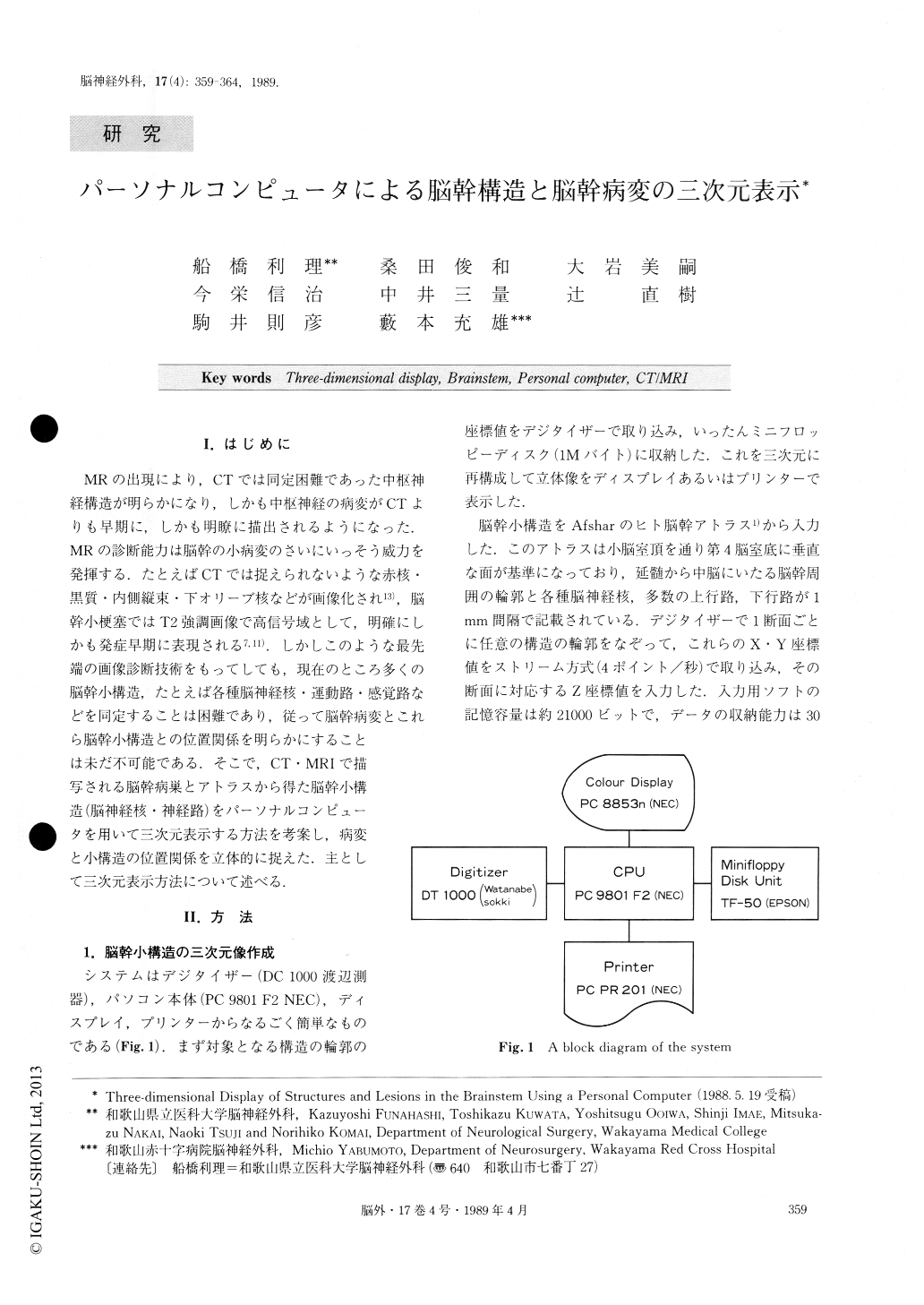
MRの出現により,CTでは同定困難であった中枢神経構造が明らかになり,しかも中枢神経の病変がCTよりも早期に,しかも明瞭に描出されるようになった.MRの診断能力は脳幹の小病変のさいにいっそう威力を発揮する.たとえばCTでは捉えられないような赤核・黒質・内側縦束・下オリーブ核などが画像化され13),脳幹小梗塞ではT2強調画像で高信号域として,明確にしかも発症早期に表現される7,11).しかしこのような最先端の画像診断技術をもってしても,現在のところ多くの脳幹小構造,たとえば各種脳神経核・運動路・感覚路などを同定することは困難であり,従って脳幹病変とこれら脳幹小構造との位置関係を明らかにすることは未だ不可能である.そこで,CT・MRIで描写される脳幹病巣とアトラスから得た脳幹小構造(脳神経核・神経路)をパーソナルコンピュータを用いて三次元表示する方法を考案し,病変と小構造の位置関係を立体的に捉えた.主として三次元表示方法について述べる.
The method of three-dimensional reconstruction of structures and lesions in the brainstem was developed by using a personal computer to visualize the affected sites in some cranial nuclei and long tracts. Outlines of brainstem structures and lesions were digitalized manu-ally by tracing the atlas of the human brainstem and CT or MR images. Four outlines of the brainstem, the medulla, lower pons, upper puns and midbrain, and the outlines of any parenchymal structures were taken at every point 2 mm in thickness. These were used as the standard visualization of the atlas.

Copyright © 1989, Igaku-Shoin Ltd. All rights reserved.


