Japanese
English
- 有料閲覧
- Abstract 文献概要
- 1ページ目 Look Inside
I.はじめに
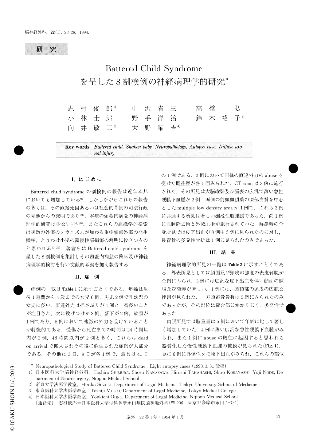
Battered child syndromeの剖検例の報告は近年本邦においても増加している9).しかしながらこれらの報告の多くは,その直接死因あるいは社会的背景の司法行政の見地からの究明であり23),本症の頭蓋内病変の神経病理学的研究は少ない5,16,20).またこれらの組織学的検索は複数の外傷のメカニズムが加わる重症頭部外傷の発生機序,とりわけ小児の瀰漫性脳損傷の解明に役立つものと思われる10,22).著者らはBattered child sYndromeを呈した8剖検例を集計しその頭蓋内病態の臨床及び神経病理学的検討を行い文献的考察を加え報告する.
The term “battered-child syndrome” was coined by Kempe in 1962. The morphology of brain lesions in abused children is rarely reported in Japan. This clinico-pathological entity in the central nervous system is characterized by retinal hemorrhages, subdural and sub-arachnoid hemorrhage. However, reports on microscopic findings of intracerebral lesion are fewer than those on macroscopic findings of scalp, skull and intracranial cavity.
This study was performed on 8 cases of battered child-ren who were autopsied. They consisted of six female and two male infants. The age ranged from one week to four years old. The causes of the injuries were shaking in four cases, throwing in three cases, dropping in two cases and strangling in one case, mostly in combination. CT scans were examined for three cases. CT scan re-vealed acute cerebral swelling and acute subdural hema-toma with interhemispheric blood clot in three cases and multiple low density area in one case. Evacuation of the subdural hematoma and external decompression was performed in one case. The survival period from injury to death was one day in four cases, and 2, 3, 9 and 41 days in the others.
In the gross anatomical findings there are many ex-coriations and bruises of the face and scalp in five cases, widespread subcutaneous hematoma in all cases and skull fracture in only two cases. The brain weight was exceedingly heavier than normal brain weight by age in five cases. In the macroscopic findings, there were marked cerebral swelling and cerebral herniation in all cases, traumatic subarachnoid hemorrhage in six cases, and thin widespread acute subdural hematoma with in-terhemispheric clot in four cases. Intraventricular hemor-rhage was found in two cases. Cerebral and gliding con-tusions were found in the temporal and bilateral parietal lobes respectively. In the microscopic findings, there were marked cerebral edema and congestion in all cases, a small hemorrhage of the bilateral basal ganglia in one case, and numerous axonal retraction balls in one case. In conclusion, we report some neuropathological find-ings for eight autopsy cases of the battered-child syn-drome. The abused children received multiple trauma from shaking, throwing, etc. In this histopathological re-port, we found some specificity of diffuse axonal injury in abused children.

Copyright © 1994, Igaku-Shoin Ltd. All rights reserved.


