Japanese
English
- 有料閲覧
- Abstract 文献概要
- 1ページ目 Look Inside
I.はじめに
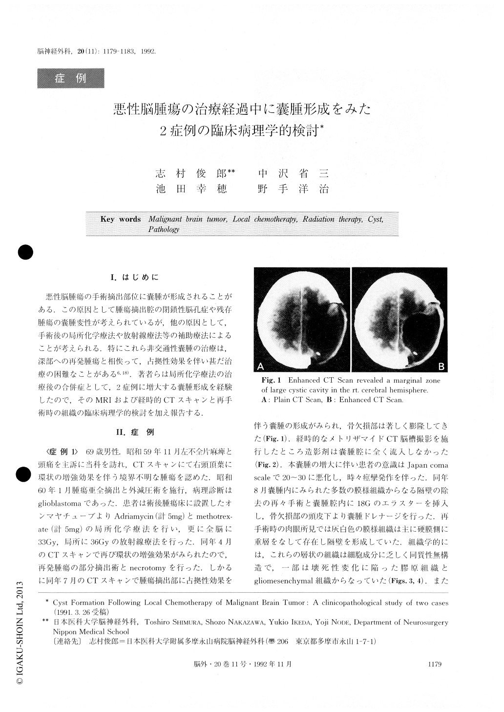
悪性脳腫瘍の手術摘出部位に嚢腫が形成されることがある.この原因として腫瘍摘出腔の閉鎖性脳孔症や残存腫瘍の嚢腫変性が考えられているが,他の原因として,手術後の局所化学療法や放射線療法等の補助療法によることが考えられる.特にこれら非交通性嚢腫の治療は,深部への再発腫瘍と相俟って,占拠性効果を伴い甚だ治療の困難なことがある6,18).著者らは局所化学療法の治療後の合併症として,2症例に増大する嚢腫形成を経験したので,そのMRIおよび経時的CTスキャンと再手術時の組織の臨床病理学的検討を加え報告する.
This paper reports clinicopathological findings concerning an enlarged bulky cyst and the tumor cavity following local administration of an anticancer agent combined with radiotherapy in two patients suffering frommalignant glioma.
Case 1: This 69 year-old man who had been diagnosed as having glioblastoma in the right parietal lobe had received local chemotherapy after the first operation. Simultaneously radiotherapy of 69 Gy in total dose was performed. At the second operation for the tumor, cyst formation was clinically confirmed and necrotomy as well as evacuation of the large cyst was performed after adjuvant therapy. The patient died at a time ten months after the first surgical operation.
Case 2 : This is the case of a 48 year-old man who was diagnosed as having gemistocytic astrocytoma in the left frontal lobe. The first surgical operation was performed and was followed by local chemotherapy as well as radiotherapy (total dose of 90 Gy in two sessions). The second surgical operation of the recurrent tumor, with necrotomy and evacuation of the large cyst were performed after adjuvant therapy. The patient expired at a time sixty-five months after the first surgical operation.
Relevant to the chemotherapy, adriamycin (ADM) (0.5mg) and methotrexate (MTX) (1mg) were administered through the Ommaya's reservoir into the tumor bed at craniotomy. The usual doses of ADM and MTX amounted to 5.0mg respectively. Through conventional CT and MRI, formation of a cyst including abundant membranous debris or septi was identified in both cases. After performing local chemotherapy and radiotherapy, excision of a recurrent tumor as well as the simultaneous evacuation of the large cyst was carried out to facilitate the efficacy of the second local chemotherapy.
The histo-pathological study of these lesions disclosed necrotic cellular debris, fibrinoid necrosis of the vascular channels and homogenous and amorphous membranous tissue which tended to form septi. In the adjacent area of this cystic necrosis, lymphocytic and macrophagic cell infiltrate, reactive proliferation of fibro-collagenous tissue component and glial reaction were observed.Conclusion:
Two cases of patients who suffered from malignant glioma have been reported. They received surgical operations twice and at the second operation, the recurrent tumors and enlarged cyst possessed fibrinous septi which were assumed to have resulted from the tissue reaction due to local chemotherapy and radiation.
Copyright © 1992, Igaku-Shoin Ltd. All rights reserved.