Japanese
English
- 有料閲覧
- Abstract 文献概要
- 1ページ目 Look Inside
I.はじめに
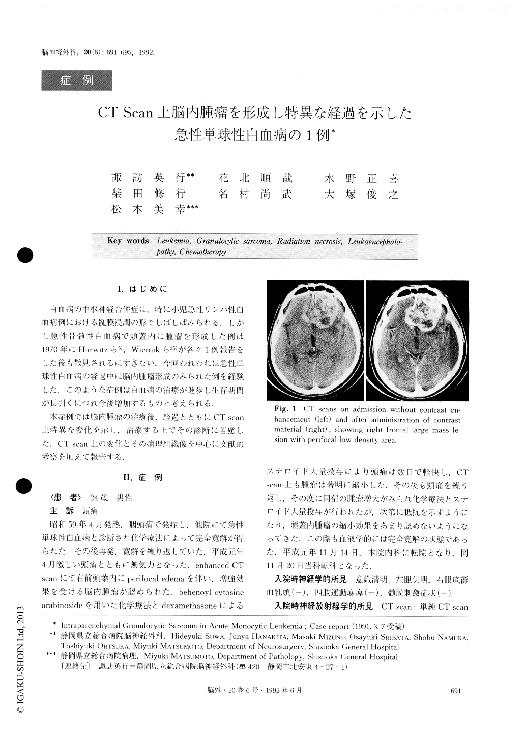
白血病の中枢神経合併症は,特に小児急性リンパ性白血病例における髄膜浸潤の形でしばしばみられる.しかし急性骨髄性白血病で頭蓋内に腫瘤を形成した例は1970年にHurwitzら5),Wiernikら25)が各々1例報告をした後も散見されるにすぎない.今回われわれは急性単球性白血病の経過中に脳内腫瘤形成のみられた例を経験した.このような症例は白血病の治療が進歩し生存期問が長引くにつれ今後増加するものと考えられる.
本症例では脳内腫瘤の治療後,経過とともにCT scan上特異な変化を示し,治療する上でその診断に苦慮した.CT scan上の変化とその病理組織像を中心に文献的考察を加えて報告する.
Granulocytic sarcoma of the parenchyma of the brain present in a patient with acute monocytic leukemia, and its unusual course during treatment, is described.
Four years after diagnosis of acute monocytic leuke-mia, a 24-year-old man developed severe headache dur-ing its remission period. The CT scan showed large in-traparencymal mass in the right frontal lobe, which was partially removed and diagnosed as granulocytic sarco-ma. Following the operation, radiation in total dose of 35.5 Gy was given to the whole brain, and there was also left intraventricular administration of methotrexate (MTX) and cytosine arabinoside (ara-C). The treat-ment resulted in the complete disappearance of the in-traparenchymal mass apart from small calcifications. Five months later, the patient redeveloped severe headache with consciousness disturbance. CT scan re-vealed marked swelling in the left cerebral hemisphere with irregular contrast-enhanced areas. The patient died of brain herniation in spite of conservative ther-apy. Photomicroscopic findings of the left cerebral hemisphere proved the presence of “disseminated leukoencephalopathy” and the absence of tumor cells. On the other hand, the right frontal lesion consisted of no tumor cells but scar tissues. This unusual feature of the CT scan in the terminal stage might be caused by combination with the effect of highly concentrated MTX in the left cerebral hemisphere because of the in-creased permeability of the ependym and the relatively high radiosensitivity in the non-affected left cerebral hemishere.

Copyright © 1992, Igaku-Shoin Ltd. All rights reserved.


