Japanese
English
- 有料閲覧
- Abstract 文献概要
- 1ページ目 Look Inside
I.はじめに
脊髄くも膜下出血は,全くも膜下出血の1%以下を占める比較的稀な疾患であるが,その症状は突然激しい背部痛,項部硬直及び頭痛などの髄膜刺激症状が主体である.しかし稀にくも膜下血腫を形成して脊髄圧迫症状を呈する場合がある.われわれは軽微な外傷を契機とし,比較的長い経路で馬尾症状を呈した脊髄くも膜下血腫の1例を経験したので,若干の文献的考察を加えて報告する.
A case of traumatic spinal subarachnoid hematoma causing compression of the cauda equina is reported here.
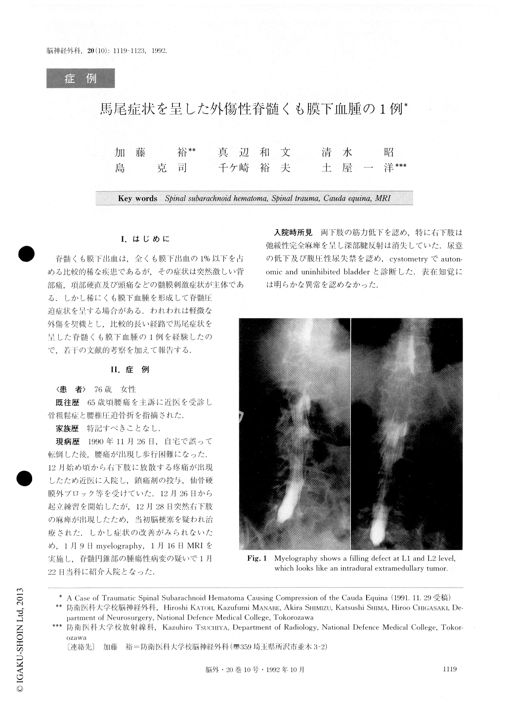
The patient, a 76 year-old woman, who had fallen down by accident 1 month before, was admitted to our hospital presenting lumbar pain radiating into her right thigh, monoplegia of the right leg and urinary inconti-nence. Myelography and metrizamide CT demonstrated a filling defect mimicking intradural extramedullary tumor at the level of Ll and L2. Magnetic resonance imagings (MRI) revealed a subacute or chronic hemato-ma compressing the conus medullaris and the cauda equina.
Operation was performed and an old hematoma, which occupied most of the spinal subarachnoid space and compressed the conus and cauda equina from right to left, was removed. No definite bleeding point was detected and no traumatic change was seen on the cord. Neither tumor nor abnormal vessel was detected. After surgery, the symptoms improved partially. On a review of the literature, we found only 4 cases of -traumatic spinal subarachnoid hematoma, all of which occupied the cervical or thoracic portion of the spine. Our case is the first report, except for the cases following lumbar spinal tap, of traumatic spinal sub-arachnoid hematoma causing compression of the cauda equina.
Though usually blood in CSF diffuses immediately, a clot may be formed when a large amount of bleeding obstructs the spinal canal. In our case, furthermore, de-formity and narrowing of the spinal canal had preceded for many years, following lumbar vertebral compressed fracture related with osteoporosis. This might have promoted the process of canal obstruction and clot formation.
Myelography and metrizamide CT indicated merely a space occupying mass in the spinal canal, but MRI con-firmed the diagnosis of the hematoma. MRI was very useful in determining the exact location and diagnosis of spinal subarachnoid hematoma.
Copyright © 1992, Igaku-Shoin Ltd. All rights reserved.