Japanese
English
- 有料閲覧
- Abstract 文献概要
- 1ページ目 Look Inside
I.はじめに
前頭蓋底脳瘤は,出生35,000)−40,000に1例で,脳瘤の1-10)%と報告されきわめて稀な先天奇形である.今回われわれは,髄膜炎にて発症しCT,MRIにて診断し開頭手術にて根治し得たtransethmoidal encephaloceleの1例を経験したので,ここに報告し考察を加える.
A case of a 3-year-old boy with transethmoidal en-cephlocele is presented.
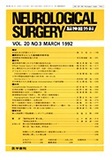
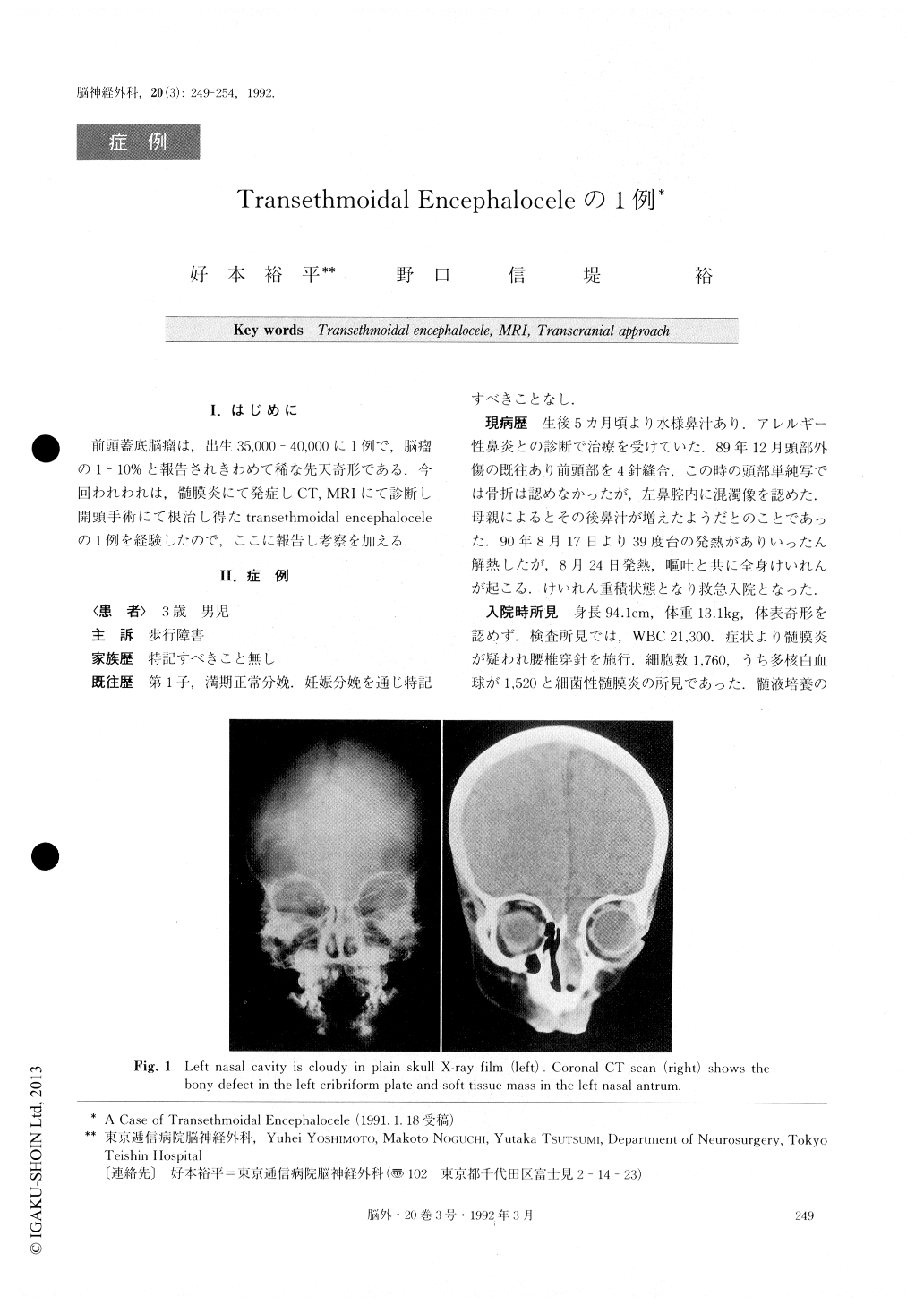
The patient was found to have bacterial meningitis, which responded well to an intravenous antibioticstheraphy. No physical anomaly was evident on ex-amination but plain skull X-ray film showed cloudiness of the left nasal antrum. Coronal CT scan disclosed a defect in the left cribriform plate and soft tissue mass in the left nasal cavity. MRI showed an anterior basal encephalocele protruding into the nasal cavity. Hypothalamic-pituitary system and the optic nerves appeared normal in the sagittal image. CSF rhinorrhea was confirmed by RI cisternography.
An operation was performed transcranially. After a left frontal craniotomy, a unilateral bony defect in the cribriform plate and protrusion of the brain was observed subfrontally. The crista galli was intact. The herniated brain substance was transected and partially removed and the bony defect plugged by temporal muscle and covered by lyofirized dura. Microscopic ex-amination of the herniated brain mass revealed gliosis and capillary proliferation. The patient recovered well and there has been no recurrence of CSF rhinorrhea or meningitis.
Basal encephalocele is a very rare congenital anoma-ly. It is reported to constitute 1 to 10% of all encepha-loceles. Incidence is estimated as 1 in every 35,000 to 40,000 live births. The anomaly is classified into two subtypes ; transethmoidal (TE) and transsphenoidal (TS) . TE rarely has associated anomalies and the pro-truded brain doesn't involve vital structures. Transsec-tion of the herniated brain and repair of the bony de-fect can be safely carried out. Most reported cases (33/ 35 cases) were operated on transcranially, all of which had good results. By contrast, most cases of TS are associated with anomalies of the face (hypertelorism, cleft lip, palate and nose), the eyes, and the brain (agenesis of corpus callosum), suggesting the pathogenesis of the condition lies in the disturbance of separation of the neuroectoderm from the ectoderm during the first 4 to 6 weeks of gestation. In this sub-type (TS), the herniated brain involves the hypothala-mic-pituitary system or optic nerves. Surgical resection is thus unacceptable because of the risks involved. Op-erative results are not satisfactory either by the trans-cranial or the transpalatal approach. CT scan is very useful to identify the bony defect of the anterior skull base and the authors would like to stress the value of MRI in identifying the content of the herniated tissue, and in helping in the decision whether to operate or not, and which approach to take.
Copyright © 1992, Igaku-Shoin Ltd. All rights reserved.