Japanese
English
- 有料閲覧
- Abstract 文献概要
- 1ページ目 Look Inside
I.はじめに
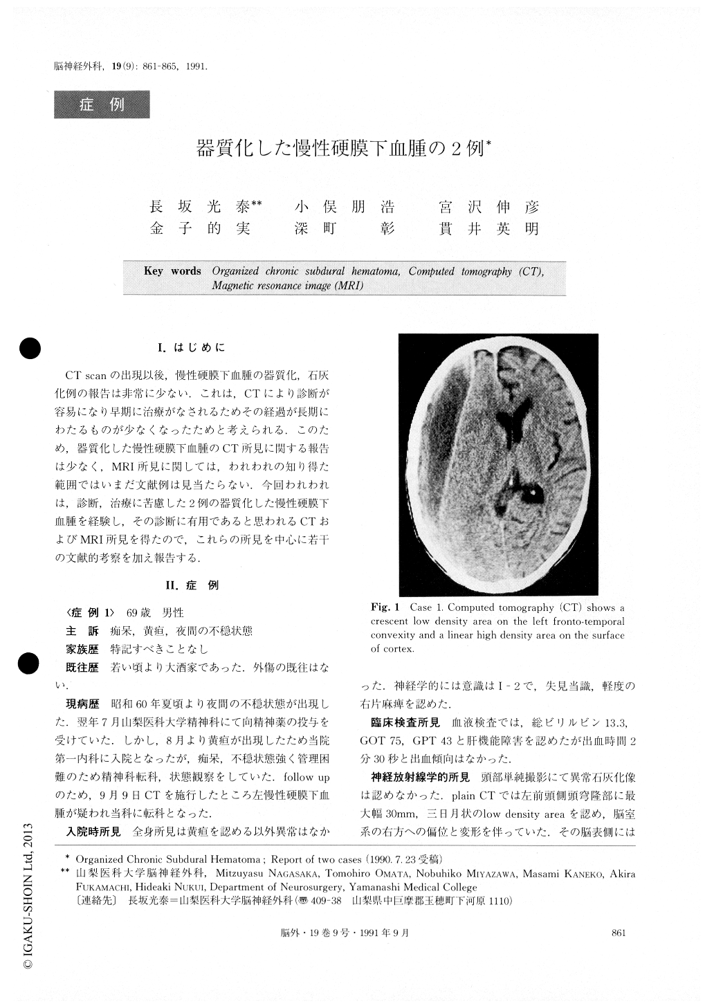
CT scanの出現以後,慢性硬膜下血腫の器質化,石灰化例の報告は非常に少ない.これは,CTにより診断が容易になり早期に治療がなされるためその経過が長期にわたるものが少なくなったためと考えられる.このため,器質化した慢性硬膜下血腫のCT所見に関する報告は少なく,MRI所見に関しては,われわれの知り得た範囲ではいまだ文献例は見当たらない.今回われわれは,診断,治療に苦慮した2例の器質化した慢性硬膜下血腫を経験し,その診断に有用であると思われるCTおよびMRI所見を得たので,これらの所見を中心に若干の文献的考察を加え報告する.
Abstract
Two cases of organized chronic subdural hematoma were presented. The first case had a one-year history of disorientation and right hemiparesis. CT scan revealed a low density area with linear high density in its medial margin, suggesting chronic subdural hematoma on the left frontal convexity. Surgery was performed expecting to remove the hematoma. There was, however, only a little fluid inside with thick membranous tissue. The second case, who has Crouzon disease, presented a one-year history of pseudobulbar palsy and tetraparesis af-ter surgery for chronic subdural hematoma and hy-drocephalus. The diagnosis of organized subdural hematoma was made at the time of reoperation which was performed expecting to remove the recurrent chro-nic subdural hematoma. Plain CT, done after admission to our hospital, showed homogenous low density area remaining in the bilateral frontal convexity. Infusion scan revealed marked enhancement of the medial mar-gin of the low density area. The lesion was demons-trated as a low intensity area by Ti-weighted magnetic resonance images (MRI). Marked enhancement was noted around the low intensity area after the infusion of Gd-DTPA. Although it is very hard to make a di-agnosis of organized chronic subdural hematoma using only the CT scan preoperatively, combination of the CT scan and MRI with Gd-DTPA enhancement seemed to be very useful for this purpose.

Copyright © 1991, Igaku-Shoin Ltd. All rights reserved.


