Japanese
English
- 有料閲覧
- Abstract 文献概要
- 1ページ目 Look Inside
I.はじめに
脳室内に発生する嚢胞性病変は頻度が少なく,側脳室では大部分がくも膜嚢胞であり,第3脳室では上衣嚢胞であることが多い.しかし神経放射線学的にそれらを鑑別することは非常に困難であり,確定診断は摘出標本の病理組織に頼ることになる14,20).
今回われわれは脳梁部分欠損症を合併した右側脳室内glio-ependymal cystの1症例を経験したので報告する.
Abstract
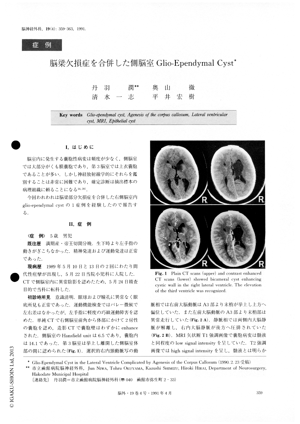
The case is that of a 5-year-old male who was admit-ted to the hospital for a further examination because of the onset of seizure observed twice. He was delivered at full term by Cesarean section, and had had impair-ment of movement of his left hand since birth. The re-sults of the first examination performed at the time of his admission to the hospital revealed mild neurological disturbance of the left hand. The CT scanning per-formed showed partial agenesis of the corpus callosum and a bicameral cyst enhancing the cystic wall extend-ing from the right anterior horn of the lateral ventricle to the body. By MRI sagittal plane, cystic masses presented low signal intensity on the T-1 weighted image, and they showed high signal intensity on the T-2 weighted im-age. The coronal plane showed that the cysts extended from the midline of the ventricle to the lateral. Cystectomy was performed using the transventricular approach. Thus communication between the cyst and lateral ventricle was made possible. The cystic wall was macroscopically white and elastically soft, and contain-ed vascular components.
Histopathologically, it consisted of 3 layers of ciliated cuboidal epithelium, glia cells and connective tissue res-pectively. We diagnosed the condition as glio-ependy-mal cyst in the lateral ventricle complicated by partial agenesis of the corpus callosum.

Copyright © 1991, Igaku-Shoin Ltd. All rights reserved.


