Japanese
English
- 有料閲覧
- Abstract 文献概要
- 1ページ目 Look Inside
I.はじめに
Meningeal gliomatosis(MG)は,悪性脳腫瘍の治療成績の向上による患者の生存期間の延長とともに,増加する傾向にあるが,その治療は非常に困難で,いまだに効果的な治療法は確立されていないといえる.このような予後不良な病態に対する治療法の研究に有用な実験モデルとして,吉田ら6)はMG modelを開発し,種々の利点を挙げている.
一方,悪性glioma髄腔内播種の成立には腫瘍細胞の遊離,着床,進展などの機転が考えられているが,その病態に関しての超微細的研究は著者らが渉猟し得た限りでは見当らない.今回著者らは吉田ら6)と同様のMGmodelを作成し,脊髄に形成された播種巣の進展様式について光顕および走査型,透過型電子顕微鏡にて観察し,若干の知見を得たので報告する.
We tried to examine the growth pattern of meningeal gliomatosis (MG) by using an experimental MG model. C6 rat glioma cells (3 × 10⁵/0.1 ml) were injected per-cutaneously into the cisterna magna of rats. Seven days after inoculation, the brains and spinal cords were re-moved and processed for morphological observation.
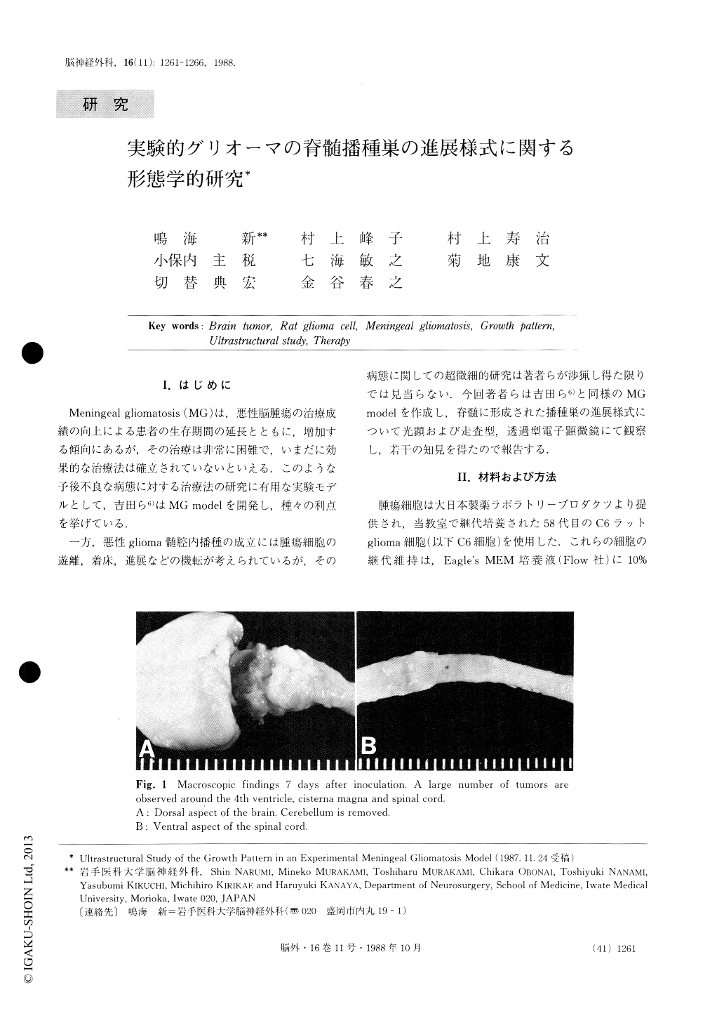
Light microscopic findings showed that numerous tumor cells had invaded the spinal cord parenchyma directly and/or via the Virchow-Robin spaces. In con-trast, a small amount of tumor cells had spread horizon-tally on the surface of the spinal cord.
Copyright © 1988, Igaku-Shoin Ltd. All rights reserved.