Japanese
English
- 有料閲覧
- Abstract 文献概要
- 1ページ目 Look Inside
I.はじめに
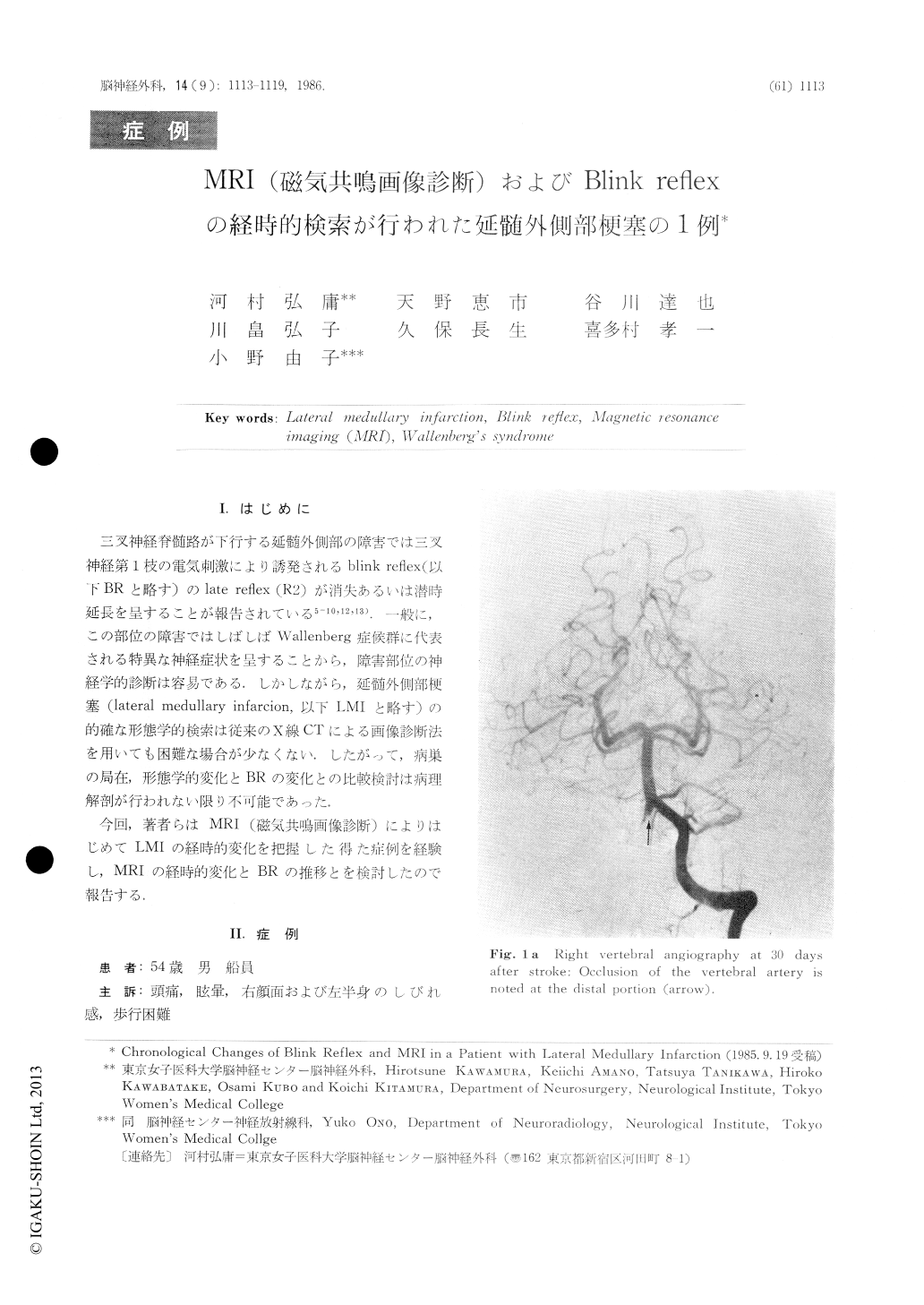
三叉神経脊髄路が下行する延髄外側部の障害では三叉神経第1枝の電気刺激により誘発されるblink reflex(以下BRと略す)のlate reflex(R2)が消失あるいは潜時延長を呈することが報告されている5-10,12,13).一般に,この部位の障害ではしばしばWallenberg症候群に代表される特異な神経症状を呈することから,障害部位の神経学的診断は容易である.しかしながら,延髄外側部梗塞(lateral medullary infarcion,以下LMIと略す)の的確な形態学的検索は従来のX線CTによる画像診断法を用いても困難な場合が少なくない.したがって,病巣の局在,形態学的変化とBRの変化との比較検討は病理解剖が行われない限り不可能であった.
今回,著者らはMRI(磁気共鳴画像診断)によりはじめてLMIの経時的変化を把握した得た症例を経験し,MRIの経時的変化とBRの推移とを検討したので報告する.
Recently, the brainstem pathways of bilateral late reflexes (R2) of electrically elicited blink reflex have been well established. An afferent delay or block of the late reflexes is closely related to a lesion of the lateral medullary portion.
The chronological alteration of blink reflex (BR) was studied to compare with radiological abnormalities on MRI in a patient with lateral medullary infarction on the right side. A diagnosis of Wallenberg syndrome was made clinically and location of the lesion was identified in detail by MRI.

Copyright © 1986, Igaku-Shoin Ltd. All rights reserved.


