Japanese
English
- 有料閲覧
- Abstract 文献概要
- 1ページ目 Look Inside
I.はじめに
頭蓋骨巨細胞腫は1936年,Geshickter8)により蝶形骨に発生した巨細胞腫の1例が報告されて以来,文献的には五十数例にすぎず,未報告例も少なくないとしても,比較的少ない疾患である,頭蓋骨での発生部位をみると,報告例では蝶形骨がその半数以上を占め,次いで側頭骨に多い,中尾18)らは1982年,本誌上に側頭骨に発生した巨細胞腫の1例を報告している.
蝶形骨に発生した巨細胞腫は,頭蓋骨巨細胞腫のなかではその頻度が多いのみならず,腫瘍の位置する解剖学的関係の複雑さのために,臨床上,特に治療上,側頭骨などの表在性の巨細胞腫とは異なった幾つかの問題を有している.われわれは最近,放射線療法が著効を呈した蝶形骨に発生した巨細胞腫を経験したので報告するとともに,焦点を蝶形骨の巨細胞腫に絞って,臨床上の若干の問題点を文献的考察を加えて論じてみたい.
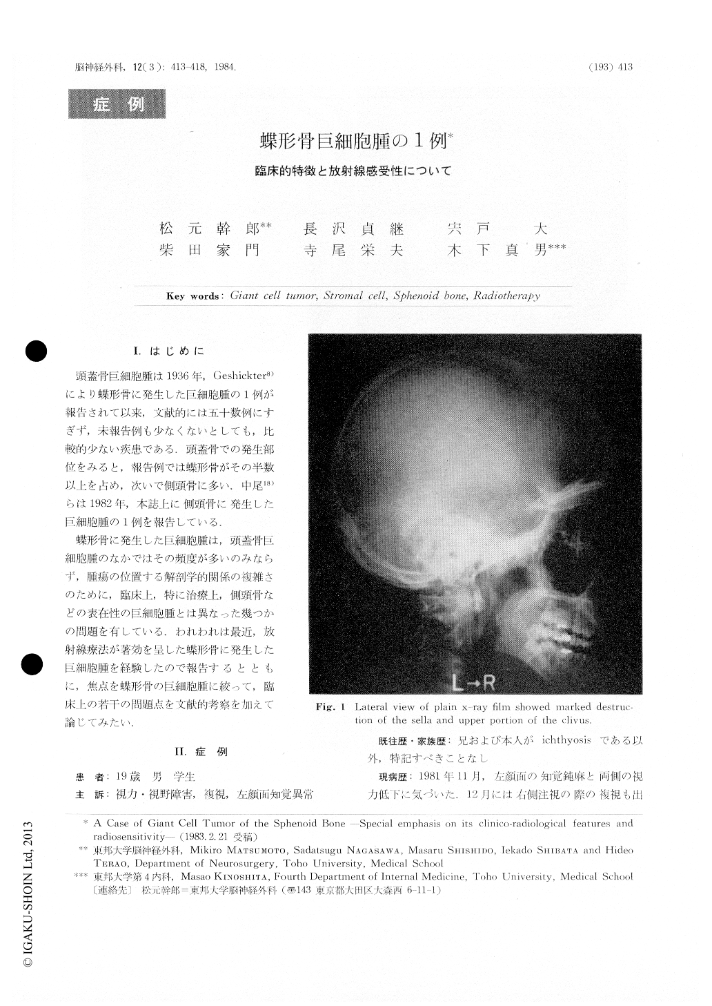
A 19-year-old man was admitted to the hospitalbecause of blurred vision, visual field defect,diplopiaand hypesthesia of the left face. Neurological ex-amination on admission revealed impairments ofthe II, III, IV, VI cranial nerves bilaterally and thefirst branch of the V nerve on the left. X-ray filmsof the skull showed a marked decalcification of thesella and upper portion of the clivus. Cerebralangiographv demonstrated a moderatedly vascularized,large tumor in the sella-clival region. The tumor wassupplied mainly by the branches of the right internalcarotid artery, which was occluded at the cavernousportion.

Copyright © 1984, Igaku-Shoin Ltd. All rights reserved.


