Japanese
English
- 有料閲覧
- Abstract 文献概要
- 1ページ目 Look Inside
I.はじめに
血管外皮26)に由来する腫瘍をStout&Murray22),Stout21)は血管外皮腫と称し,その半数は筋や骨格に発生する,髄膜に発生した血管外皮腫についてはBegg&Garret4)が1954年に初めて報告した.このものは髄膜に付着し,肉眼的に髄膜腫と差がなく,Baileyら3),Russellら18)は血管外皮腫を悪性徴候のなし血管の豊富な髄膜腫と天幕上の良性血管腫を一括して血管芽細胞性髄膜腫(angioblastic meningioma)としている,Ker-nohan & Uihlein10)は髄膜に発生した血管外皮腫において多数の核分裂像と豊富な格子線維網の存在,および再発傾向から髄膜腫と区別して肉腫の一種としている.Simpson19)も髄膜に発生した血管外皮腫を未分化肉腫と呼んでいる,WHO分類(World Health Organiza-tion,1979)27)によると血管外皮腫を髄膜腫の一型とし,その悪性度はgrade IIとしている.以上のように血管外皮腫の位置づけに見解の一致をみていない.
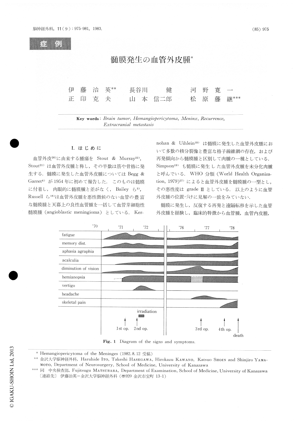
A 44-year-old farmer complained blurred vision anddisturbance of recent memory. During his drivingcar traffic accident happened due to his right homo-nymous hemianopsia. On the 1st admission, neuro-logical examination revealed choked disc (1 D.),hemi-anopsia, memory disturbance, clyscalculia, dyslexiaanddysgraphia.
The angiograms showed feeding arteries from leftmiddle cerebral artery and posterior cerebralartery.
Tumor vessels looked like cork-screw in thearterialphase and homogeneous tumor shadow was depictedin late venous phase.

Copyright © 1983, Igaku-Shoin Ltd. All rights reserved.


