Japanese
English
- 有料閲覧
- Abstract 文献概要
- 1ページ目 Look Inside
I.はじめに
大脳縦裂部硬膜下血腫の報告例は,CTの普及した現在なお少なく,文献上十数例の報告をみるにすぎない1,4,6,8-10).大部分の症例は頭部外傷に起因しており,血液疾患に伴って発生したとする報告は見当らないようである.また,大脳縦裂部以外の部位に同時に硬膜下血腫がみられることも成人例では極めて少ないようである3).
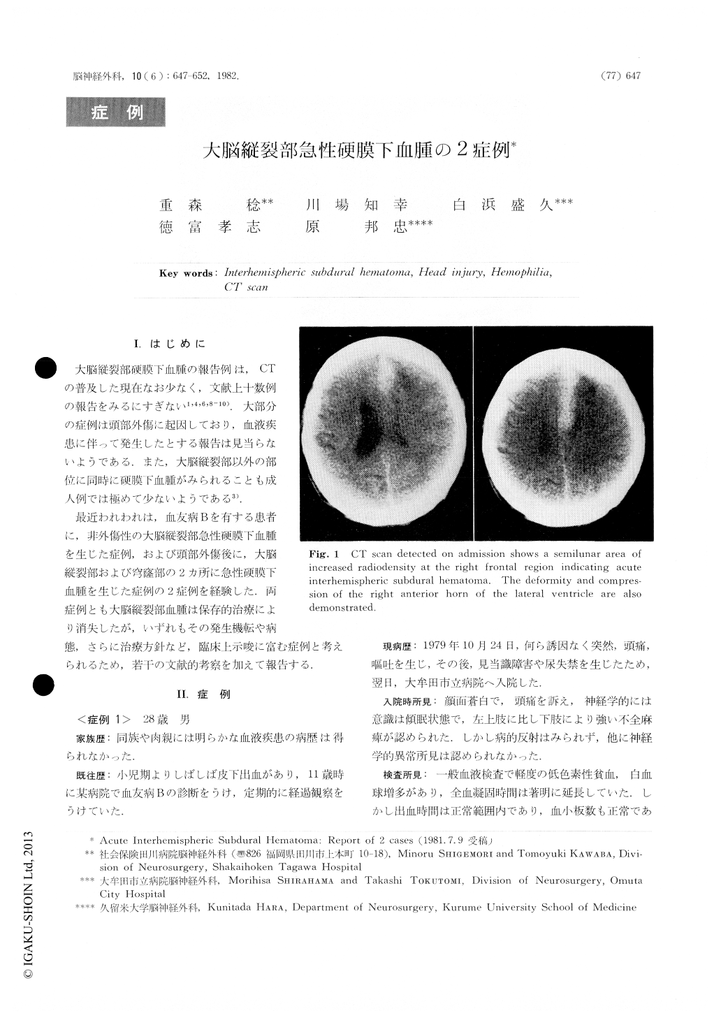
最近われわれは,血友病Bを有する患者に,非外傷性の大脳縦裂部急性硬膜下血腫を生じた症例,および頭部外傷後に,大脳縦裂部および穹窿部の2ヵ所に急性硬膜下血腫を生じた症例の2症例を経験した.両症例とも大脳縦裂部血腫は保存的治療により消失したが,いずれもその発生機転や病態,さらに治療方針など,臨床上示唆に富む症例と考えられるため,若干の文献的考察を加えて報告する.
Two cases of acute interhemispheric subduralhematomasdeveloped in a hemophiliac and following head injurywerereported, with special reference to the possiblemechanismsof production of these hematomas and treatment.
Case 1: A 28-year-old man with known hemophilia Bcomplained of severe headache and vomiting whichdev-eloped without previous head trauma and admitted toOmuta City Hospital on October 25, 1979. Onadmission,he was lethargic and mild hemiparesis on the leftside,marked in the lower extremity was noted. CI scanrevealeda characteristic feature of interhemisphericsubdural hema-toma in the right frontal region.

Copyright © 1982, Igaku-Shoin Ltd. All rights reserved.


