Japanese
English
- 有料閲覧
- Abstract 文献概要
- 1ページ目 Look Inside
I.はじめに
硬膜下膿瘍は全頭蓋内感染症の13-20%16,24)を占め,臨床的にも既に各方向から検討されてきた1,3,6,16,24),しかし,大脳縦裂膿瘍はその報告も少なく,病態の把握および治療は必ずしも容易でなく,他部位の硬膜下膿瘍と様相を異にするといわれている10,12,13,24).硬膜下膿瘍の診断は従来,脳血管撮影,isotope brain scanに頼る以外になかった4,5,19,20,22,23)が,CT scanの導入以後,他検査では得られぬ,より多くの正確な情報が得られ,侵襲も少なく,手術適応の決定,術後の追跡観察に最も有用であることが指摘されている7,8,9).しかし,大脳縦裂膿瘍に関するCT scanの報告は少なく7,8,11,15,17,21).本邦では和賀の1例のみである21).われわれは最近,CT scanで大脳縦裂膿瘍を主とした多発性硬膜下膿瘍の1例を診断し,手術によってこれを確認,治癒せしめ,CT scanで術前術後の経過観察をなしえた.
Multiple subdural abscesses, including one in the interhemispheric fissure were well diagnosed by CT scan and were successfully treated by surgery which confirmed the abscesses to have been isolated from each other.
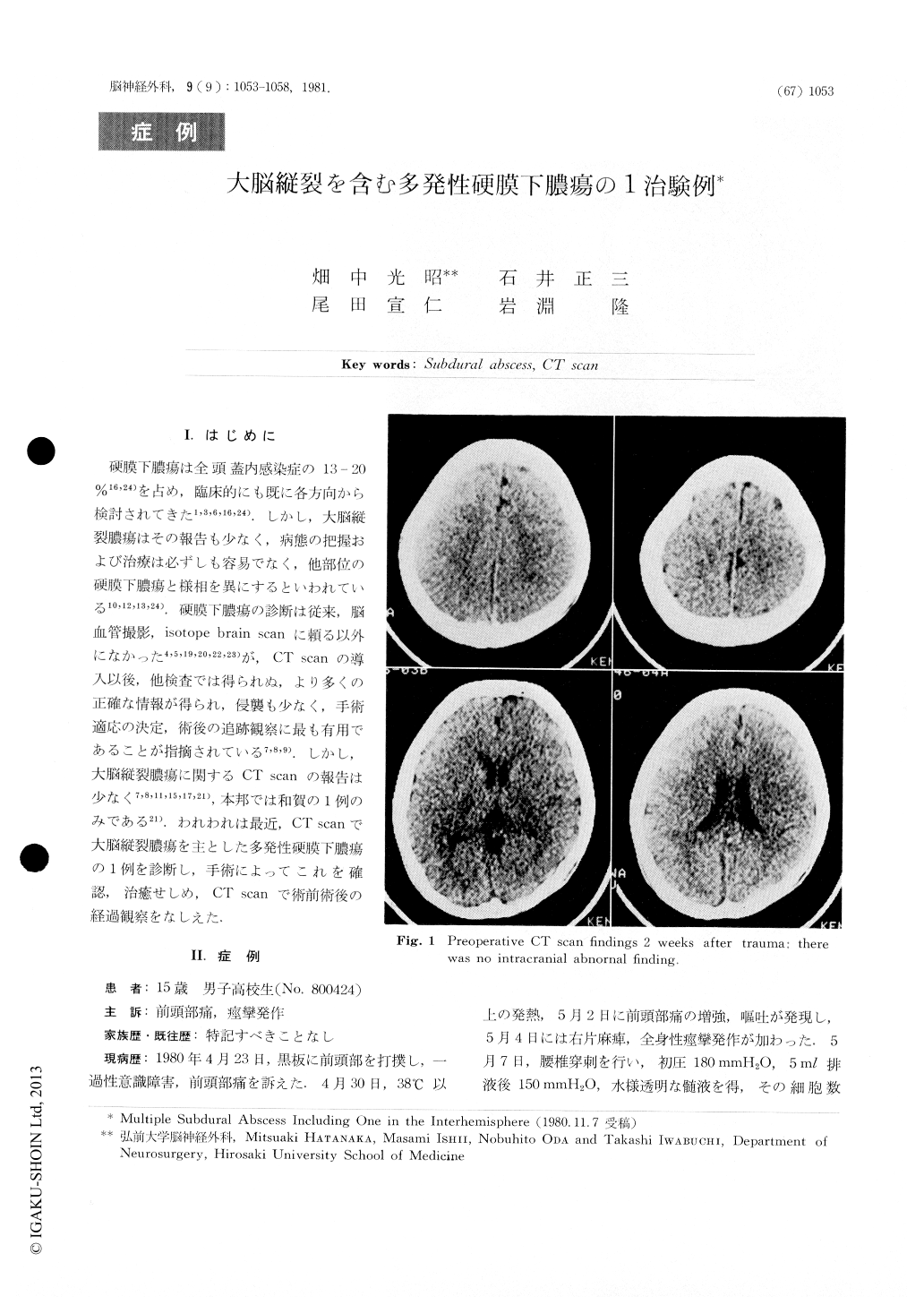
A 15-year-old boy started to complain of fever, headache and vomiting 7 days after a frontal contusion, but a CT scan showed only high density in the frontal nasal sinus. On the 11 th post-traumatic day, he had an epileptic seizure followed by right hemiparesis and motor aphasia, and was admitted to our clinic.

Copyright © 1981, Igaku-Shoin Ltd. All rights reserved.


