Japanese
English
- 有料閲覧
- Abstract 文献概要
- 1ページ目 Look Inside
I.はじめに
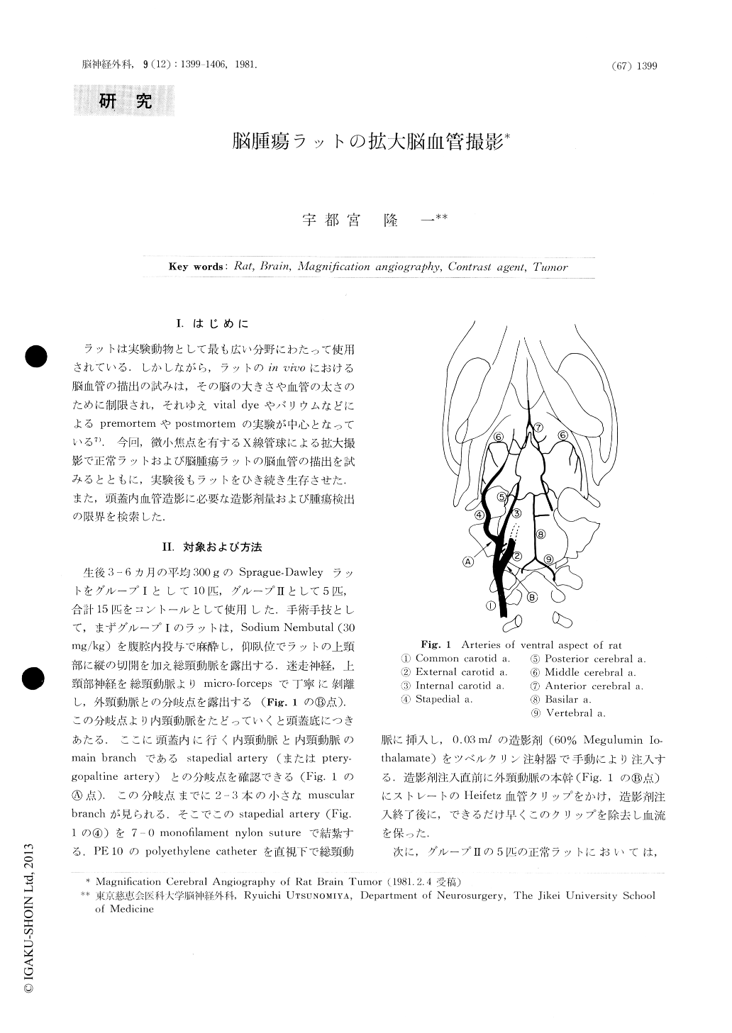
ラットは実験動物として最も広い分野にわたって使用されている.しかしながら,ラットのin vivoにおける脳血管の描出の試みは,その脳の大きさや血管の太さのために制限され,それゆえvital dyeやバリウムなどによるpremortemやpostmortemの実験が中心となっている7).今回,微小焦点を有するX線管球による拡大撮影で正常ラットおよび脳腫瘍ラットの脳血管の描出を試みるとともに,実験後もラットをひき続き生存させた.また,頭蓋内血管造影に必要な造影剤量および腫瘍検出の限界を検索した.
Rats have been widely used as laboratory animals because they are durable and inexpensive. Radiologic attempts to study their intracranial circulation have been limited to either premortem or postmortem perfusion studies using substances such as vital dyes and micropaque. A technique to opacify the intracranial circulation of rats in vivo will be described. The optimum dose of contrast agent needed to visualize the intracranial vessels will be suggested.
Fifteen normal rats and ten tumor rats induced by ethylnitrosourea (50mg/kg) were used in this study. The animals were anesthetized with sodium nembutal (20-30 mg/kg).

Copyright © 1981, Igaku-Shoin Ltd. All rights reserved.


