Japanese
English
- 有料閲覧
- Abstract 文献概要
- 1ページ目 Look Inside
抄録
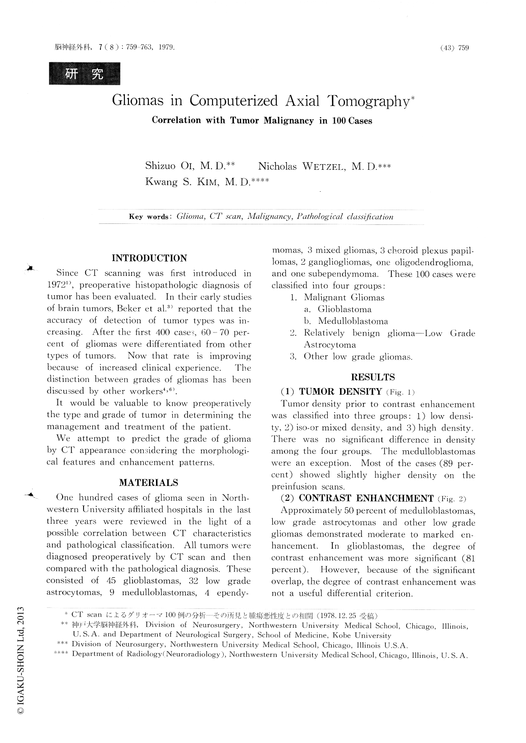
CT scanの臨床への応用がなされて以来,従来の神経放射線学的診断法は大きく変化した.それとともに脳腫瘍における診断のアプローチもCT scanを中心として変化してきた.しかしながら,CT scanにおける腫瘍の組織学的診断はまだ確立されたものではなく,未だ漠然とした鑑別診断の域を脱し得ない.その理由として,1つはCT scanにおける良性及び悪性腫瘍の所見にoverlapしたものが多いこと,そしてもう1つの理由として,各腫瘍の所見でいわゆる"典型像"に関しては一応の見解の一致がみられるが,いくつかの例外があり,今後さらに症例を重ねる必要があること,などがあげられよう.
我々は,CT scanによる脳腫瘍の診断に関していくつかの観点から検討してきたが,今回特にその悪性度の診断について,病理学的に診断さらに分類されたグリオーマの100例のCT scanの所見を分析した.これらの結果より,グリオーマの悪性度を高率に暗示するものとして,1)post infusion homogenecity(central low densityのあるものでは,壁の厚さと不規則性),2)peritumoral edema,3)腫瘍の辺縁の不規則性,があげられる.また悪性度と相関性の乏しいものとして 1)preinfusion density,2)contrast enhancelnent,があげられ良性・悪性にoverlapする所見として注意する必要があると思われる.
INTRODUCTION
Since CT scanning was first introduced in 19721), preoperative histopathologic diagnosis of tumor has been evaluated. In their early studies of brain tumors, Beker et al.3) reported that the accuracy of detection of tumor types was increasing. After the first 400 cases, 60 - 70 percent of gliomas were differentiated from other types of tumors. Now that rate is improving because of increased clinical experience. The distinction between grades of gliomas has been discussed by other workers4,6).
It would be valuable to know preoperatively the type and grade of tumor in determining the management and treatment of the patient.

Copyright © 1979, Igaku-Shoin Ltd. All rights reserved.


