Japanese
English
- 有料閲覧
- Abstract 文献概要
- 1ページ目 Look Inside
- 参考文献 Reference
Ⅰ.はじめに
内視鏡下の経蝶形骨洞手術が普及してきた.内視鏡手術の最大の特徴は,広く明るい手術野が得られることである.小さな鼻腔粘膜の入り口から手術可能であり,低侵襲性を狙ったendoscopic pituitary surgeryが確立した4,9,10).一方,鞍外部の視野が広く十分に得られることから,鞍外病変への拡大手術も行われている.これは拡大蝶形骨洞法,さらには,endoscopic skull base surgeryとして発展しつつある2,7,8,13).
筆者らは,上記の内視鏡の特徴を生かして,トルコ鞍外病変にも積極的に経鼻的内視鏡手術を行っている.開創器を使わないことで,見えるけれど器具が届かない内視鏡手術の欠点を補える10,11,12).
今回は,その手術施行症例を紹介し,本法の将来を展望する.
Background and Objectives
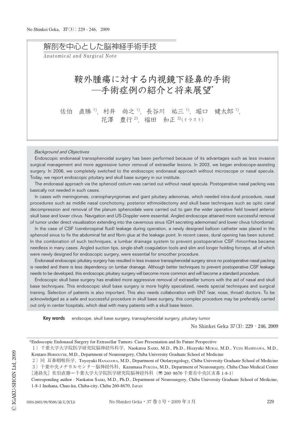
Endoscopic endonasal transsphenoidal surgery has been performed because of its advantages such as less invasive surgical management and more aggressive tumor removal of extrasellar lesions. In 2003, we began endoscope-assisting surgery. In 2006, we completely switched to the endoscopic endonasal approach without microscope or nasal specula. Today, we report endoscopic pituitary and skull base surgery in our institute.
The endonasal approach via the sphenoid ostium was carried out without nasal specula. Postoperative nasal packing was basically not needed in such cases.
In cases with meningiomas, craniopharyngiomas and giant pituitary adenomas, which needed intra-dural procedure, nasal procedures such as middle nasal conchotomy, posterior ethmoidectomy and skull base techniques such as optic canal decompression and removal of the planum sphenoidale were carried out to gain the wider operative field toward anterior skull base and lower clivus. Navigation and US-Doppler were essential. Angled endoscope attained more successful removal of tumor under direct visualization extending into the cavernous sinus (GH secreting ademomas) and lower clivus (chordoma).
In the case of CSF (cerebrospinal fluid) leakage during operation, a newly designed balloon catheter was placed in the sphenoid sinus to fix the abdominal fat and fibrin glue at the leakage point. In recent cases, dural opening has been sutured. In the combination of such techniques, a lumbar drainage system to prevent postoperative CSF rhinorrhea became needless in many cases. Angled suction tips, single-shaft coagulation tools and slim and longer holding forceps, all of which were newly designed for endoscopic surgery, were essential for smoother procedure.
Endonasal endoscopic pituitary surgery has resulted in less invasive transsphenoidal surgery since no postoperative nasal packing is needed and there is less dependency on lumbar drainage. Although better techniques to prevent postoperative CSF leakage needs to be developed, this endoscopic pituitary surgery will become more common and will become a standard procedure.
Endoscopic skull base surgery has enabled more aggressive removal of extrasellar tumors with the aid of nasal and skull base techniques. This endoscopic skull base surgery is more highly specialized, needs special techniques and surgical training. Selection of patients is also important. This also needs collaboration with ENT (ear, nose, throat) doctors. To be acknowledged as a safe and successful procedure in skull base surgery, this complex procedure may be preferably carried out only in center hospitals, which deal with many patients with a skull base lesion.

Copyright © 2009, Igaku-Shoin Ltd. All rights reserved.


