Japanese
English
- 有料閲覧
- Abstract 文献概要
- 1ページ目 Look Inside
- 参考文献 Reference
Ⅰ.はじめに
脳腫瘍の定性的評価として201Thallium-chloride single photon computed tomography(Tl-SPECT)は,良性・悪性の鑑別診断や悪性度の不均一な腫瘍での活動性の高い部位の同定などに有用で,さらに各種の治療効果判定,残存腫瘍や局所再発の有無,放射線壊死との鑑別,化学療法の経過中における効果予測などにも有用との報告がある2,6).一方でTl-SPECTは空間分解能が低く定量性に難点があった.Tl-SPECTの定量指標(Tl値)として,tumor uptake(T)とnormal uptake(N)からの比でearly ratio(ER)=early T/N,delayed ratio(DR)=delayed T/Nがあり,さらにretention index(RI)=DR/ERあるいはmodified RI=(DR-ER)/ERなどが用いられ,脳腫瘍の良性・悪性の判定基準として報告されている3,8).
近年,SPECT画像での空間分解能向上とともに統計的手法や他のモダリティとの融合による総合的評価が行われ,特にmagnetic resonance imaging(MRI)とSPECT画像をfusionする画像ソフトが普及している7).そのようなMR fusion画像ソフトにTl用に開発されたBrain Easy Analysis Tool for 201Tl(BEAT-TL®“ビートル”;富士フィルムRIファーマ㈱,東京)があるが,このソフトを用いたTl-SPECTの定量性の報告は未だ十分でなく,脳腫瘍診断でのMR fusion画像によるTl-SPECT画像の定量性を再検討しなければならないと思われた.
今回,われわれは,BEAT-TL®が融合による診断情報の増加および関心領域(region of interest: ROI)の設定の自由度や指標算出の簡便性の点で従来のものより優れ,定量性や再現性が高いことの認識から,MR fusion Tl-SPECT(MR-F群)とnon-fusion Tl-SPECT(NF群)を比較することで,MR fusion Tl-SPECT画像の有用性および定量性について検討した.
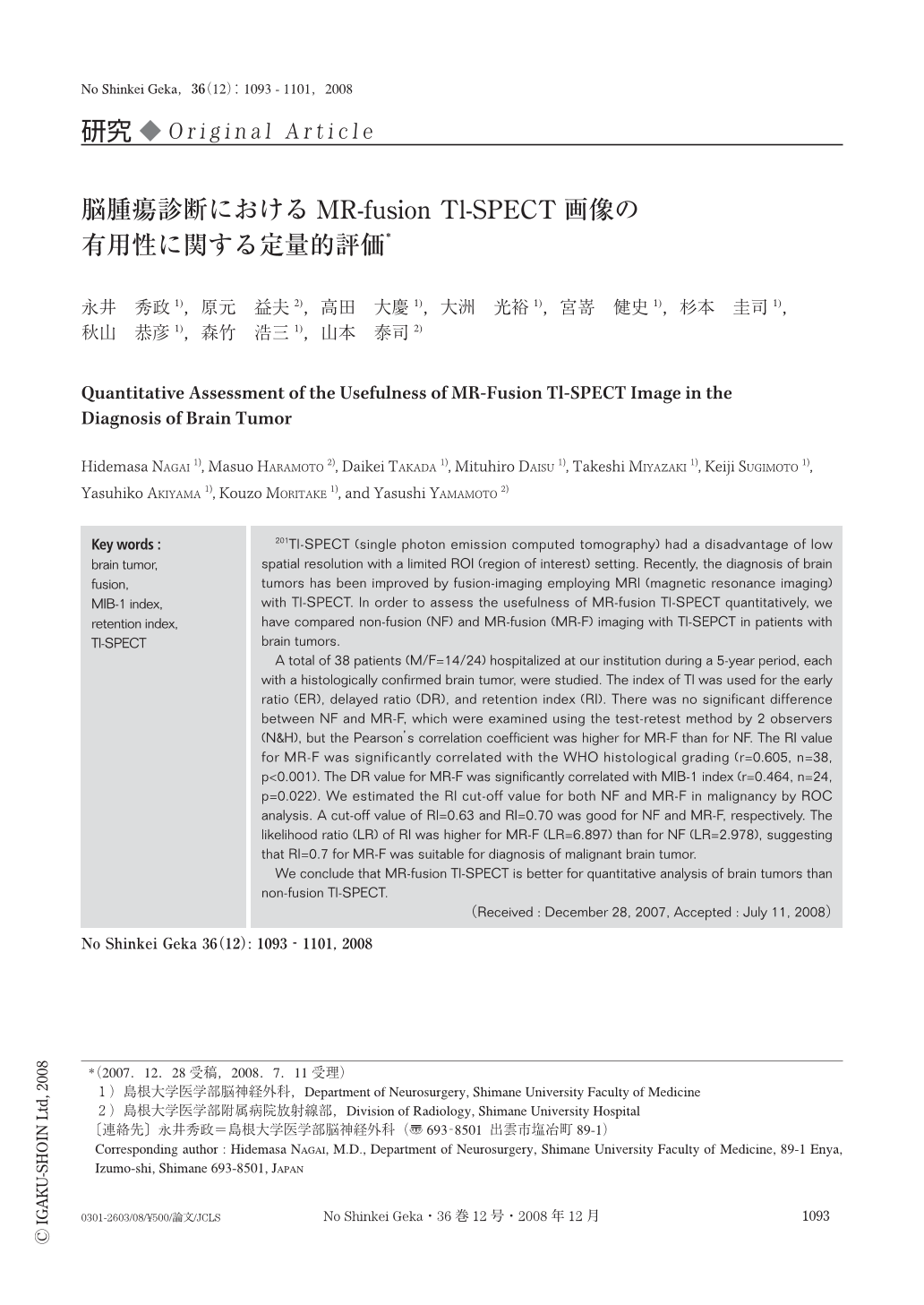
201Tl-SPECT (single photon emission computed tomography) had a disadvantage of low spatial resolution with a limited ROI (region of interest) setting. Recently, the diagnosis of brain tumors has been improved by fusion-imaging employing MRI (magnetic resonance imaging) with Tl-SPECT. In order to assess the usefulness of MR-fusion Tl-SPECT quantitatively, we have compared non-fusion (NF) and MR-fusion (MR-F) imaging with Tl-SEPCT in patients with brain tumors.
A total of 38 patients (M/F=14/24) hospitalized at our institution during a 5-year period, each with a histologically confirmed brain tumor, were studied. The index of Tl was used for the early ratio (ER), delayed ratio (DR), and retention index (RI). There was no significant difference between NF and MR-F, which were examined using the test-retest method by 2 observers (N&H), but the Pearson's correlation coefficient was higher for MR-F than for NF. The RI value for MR-F was significantly correlated with the WHO histological grading (r=0.605, n=38, p<0.001). The DR value for MR-F was significantly correlated with MIB-1 index (r=0.464, n=24, p=0.022). We estimated the RI cut-off value for both NF and MR-F in malignancy by ROC analysis. A cut-off value of RI=0.63 and RI=0.70 was good for NF and MR-F, respectively. The likelihood ratio (LR) of RI was higher for MR-F (LR=6.897) than for NF (LR=2.978), suggesting that RI=0.7 for MR-F was suitable for diagnosis of malignant brain tumor.
We conclude that MR-fusion Tl-SPECT is better for quantitative analysis of brain tumors than non-fusion Tl-SPECT.

Copyright © 2008, Igaku-Shoin Ltd. All rights reserved.


