Japanese
English
- 有料閲覧
- Abstract 文献概要
- 1ページ目 Look Inside
- 参考文献 Reference
Ⅰ.は じ め に
出生直後の新生児において産瘤や頭血腫による頭皮の膨隆はよくみられるが,そのほとんどが数週間で自然に吸収され消滅する.一方,頭血腫では残存血腫周囲が骨化し,頭蓋変形を来すことは知られているが,頭蓋形成を必要とするほど大きなものは稀である2,4,5,6,12).
われわれは頭血腫の骨化により著しい頭蓋変形を来した小児例に対し,吸収性骨プレートを用いた頭蓋形成を行った症例を経験したので,文献的考察を加え報告する.
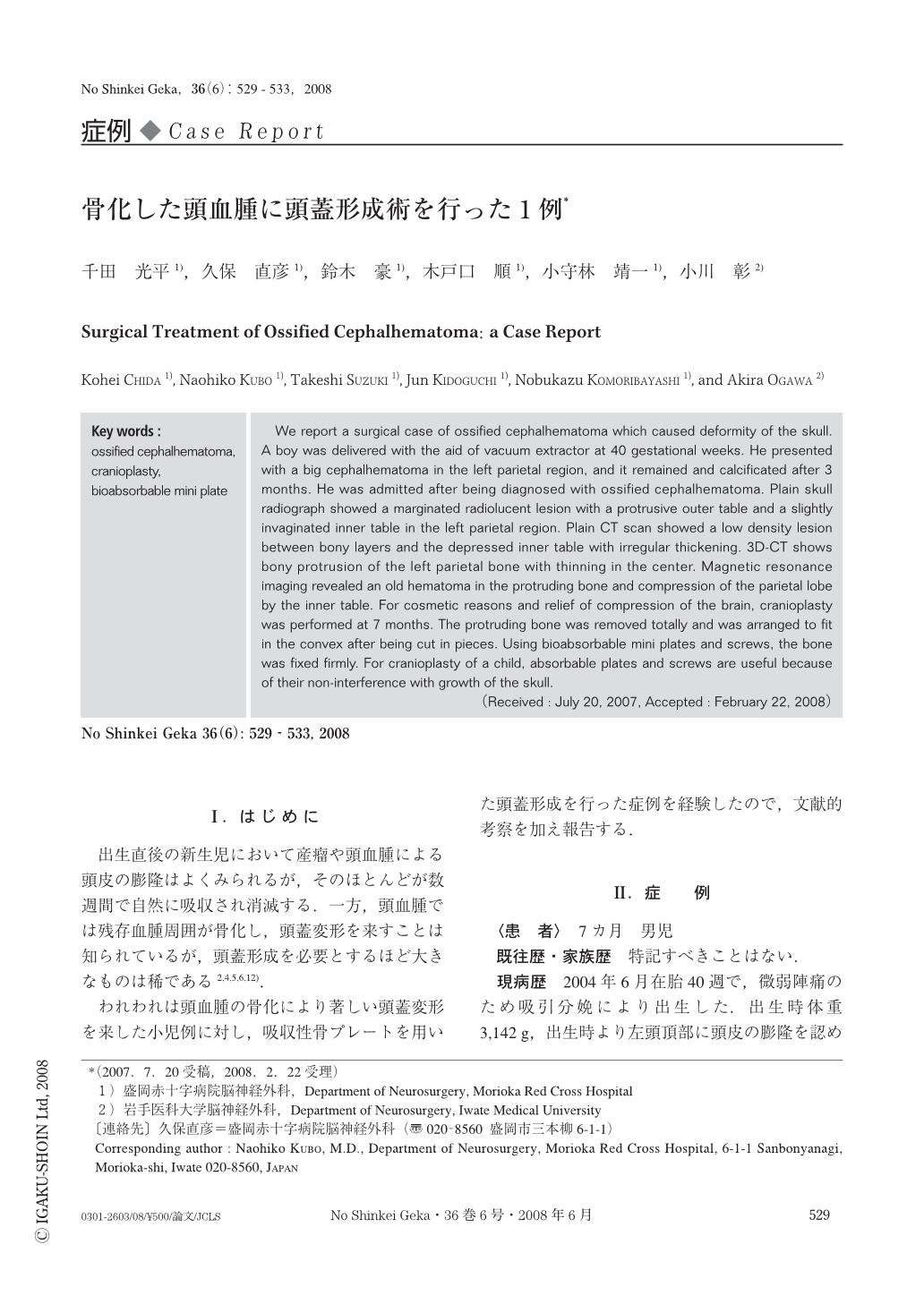
We report a surgical case of ossified cephalhematoma which caused deformity of the skull. A boy was delivered with the aid of vacuum extractor at 40 gestational weeks. He presented with a big cephalhematoma in the left parietal region, and it remained and calcificated after 3 months. He was admitted after being diagnosed with ossified cephalhematoma. Plain skull radiograph showed a marginated radiolucent lesion with a protrusive outer table and a slightly invaginated inner table in the left parietal region. Plain CT scan showed a low density lesion between bony layers and the depressed inner table with irregular thickening. 3D-CT shows bony protrusion of the left parietal bone with thinning in the center. Magnetic resonance imaging revealed an old hematoma in the protruding bone and compression of the parietal lobe by the inner table. For cosmetic reasons and relief of compression of the brain, cranioplasty was performed at 7 months. The protruding bone was removed totally and was arranged to fit in the convex after being cut in pieces. Using bioabsorbable mini plates and screws, the bone was fixed firmly. For cranioplasty of a child, absorbable plates and screws are useful because of their non-interference with growth of the skull.

Copyright © 2008, Igaku-Shoin Ltd. All rights reserved.


