Japanese
English
- 有料閲覧
- Abstract 文献概要
- 1ページ目 Look Inside
- 参考文献 Reference
Ⅰ.はじめに
基底核の石灰化はCTが開発されて以降,成人の数%とそれほど珍しいものではない1,3).しかし,圧倒的に多いのは40歳以上での淡蒼球石灰化であり,尾状核・傍側脳室部の石灰化は極めて稀である1,3).今回,われわれは基底核の石灰化病変を呈した20歳男性のKallmann症候群の症例を経験したので,文献的考察を加え報告する.
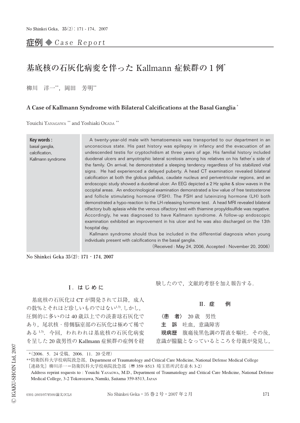
A twenty-year-old male with hematoemesis was transported to our department in an unconscious state. His past history was epilepsy in infancy and the evacuation of an undescended testis for cryptochidism at three years of age. His familial history included duodenal ulcers and amyotrophic lateral screlosis among his relatives on his father's side of the family. On arrival, he demonstrated a sleeping tendency regardless of his stabilized vital signs. He had experienced a delayed puberty. A head CT examination revealed bilateral calcification at both the globus pallidus, caudate nucleus and periventricular regions, and an endoscopic study showed a duodenal ulcer. An EEG depicted a 2Hz spike & slow waves in the occipital areas. An endocrinological examination demonstrated a low value of free testosterone and follicle stimulating hormone (FSH). The FSH and luteinizing hormone (LH) both demonstrated a hypo-reaction to the LH-releasing hormone test. A head MRI revealed bilateral olfactory bulb aplasia while the venous olfactory test with thiamine propyldisulfide was negative. Accordingly, he was diagnosed to have Kallmann syndrome. A follow-up endoscopic examination exhibited an improvement in his ulcer and he was also discharged on the 13th hospital day.
Kallmann syndrome should thus be included in the differential diagnosis when young individuals present with calcifications in the basal ganglia.

Copyright © 2007, Igaku-Shoin Ltd. All rights reserved.


