Japanese
English
- 有料閲覧
- Abstract 文献概要
- 1ページ目 Look Inside
I.緒言
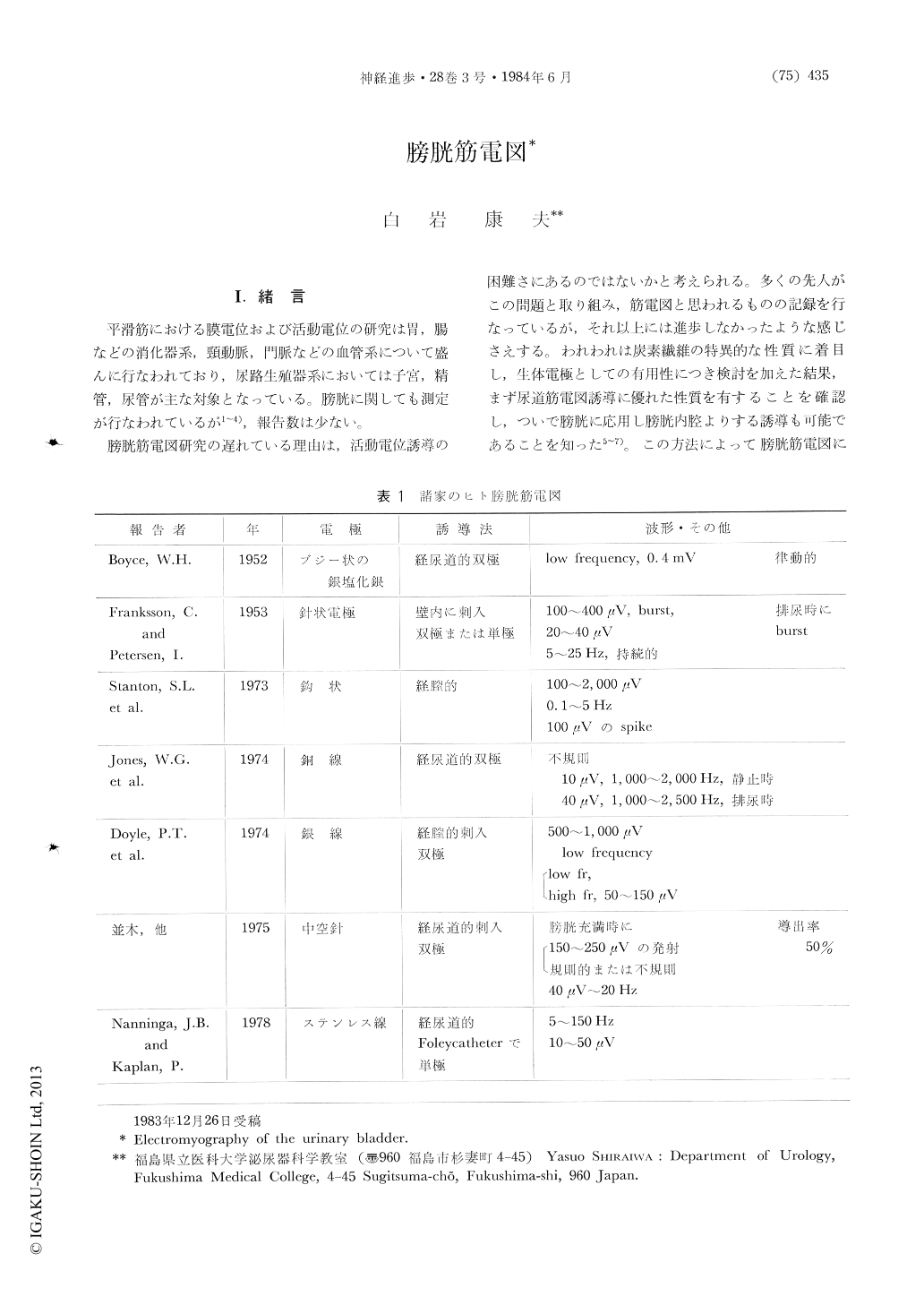
平滑筋における膜電位および活動電位の研究は胃,腸などの消化器系,頸動脈,門脈などの血管系について盛んに行なわれており,尿路生殖器系においては子宮,精管,尿管が主な対象となっている。膀胱に関しても測定が行なわれているが1〜4),報告数は少ない。
膀胱筋電図研究の遅れている理由は,活動電位誘導の困難さにあるのではないかと考えられる。多くの先人がこの問題と取り組み,筋電図と思われるものの記録を行なっているが,それ以上には進歩しなかったような感じさえする。われわれは炭素繊維の特異的な性質に着目し,生体電極としての有用性につき検討を加えた結果,まず尿道筋電図誘導に優れた性質を有することを確認し,ついで膀胱に応用し膀胱内腔よりする誘導も可能であることを知った5〜7)。この方法によって膀胱筋電図について基礎的な検討を行ない,さらに臨床応用を試み,ある程度の成果を上げることができたので,それらの成績を中心とした膀胱筋電図の概説を行なってみたい。
Abstract
The paper describes the use of carbonfibers, an exellent electrode in urinary bladder electro-myography. A bipolar electrode, made from felt-like carbonfibers, 2 mm in diameter, may serve as an electromyographic lead from the serosal surface of the dog urinary bladder or from the mucosal surface transurethrally. The latter technique may be applied clinically. Experimentally, the dog urinary bladder was exposed at laparotomy and electromyographic leads were attached to the serosal surface. With the induction of bladder contraction by the use of carbachol, a burst of spikes up to 30 μV appeared.

Copyright © 1984, Igaku-Shoin Ltd. All rights reserved.


