Japanese
English
- 有料閲覧
- Abstract 文献概要
- 1ページ目 Look Inside
はじめに
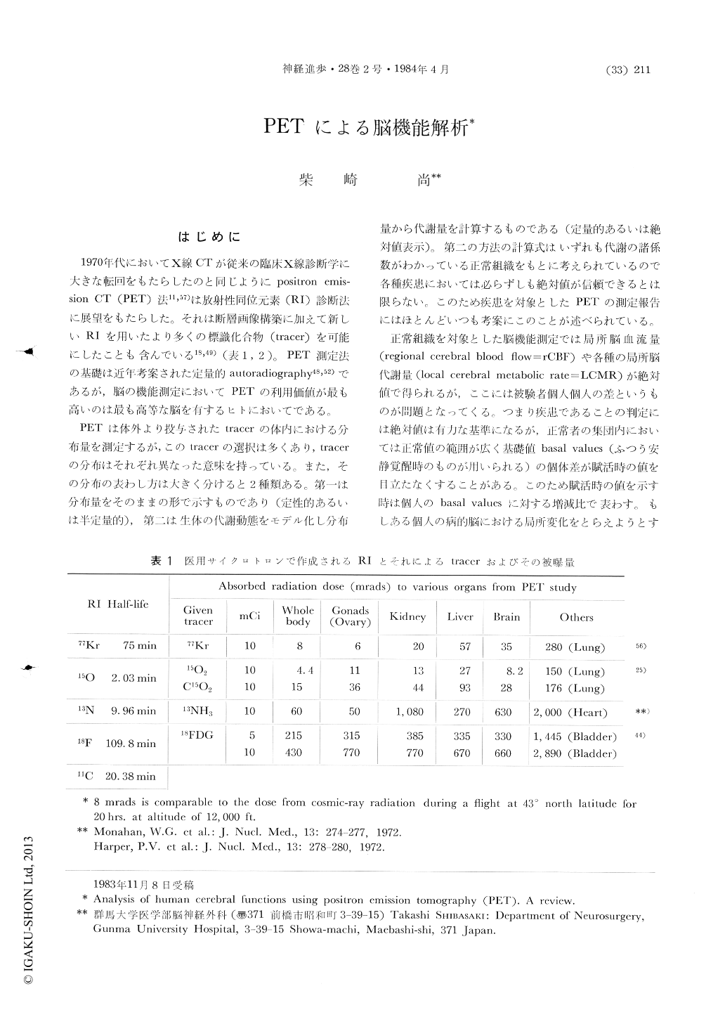
1970年代においてX線CTが従来の臨床X線診断学に大きな転回をもたらしたのと同じようにpositron emission CT(PET)法11,57)は放射性同位元素(RI)診断法に展望をもたらした。それは断層画像構築に加えて新しいRIを用いたより多くの標識化合物(tracer)を可能にしたことも含んでいる18,49)(表1,2)。PET測定法の基礎は近年考案された定量的autoradiography48,52)であるが,脳の機能測定においてPETの利用価値が最も高いのは最も高等な脳を有するヒトにおいてである。
PETは体外より投与されたtracerの体内における分布量を測定するが,このtracerの選択は多くあり,tracerの分布はそれぞれ異なった意味を持っている。また,その分布の表わし方は大きく分けると2種類ある。第一は分布量をそのままの形で示すものであり(定性的あるいは半定量的),第二は生体の代謝動態をモデル化し分布量から代謝量を計算するものである(定量的あるいは絶対値表示)。第二の方法の計算式はいずれも代謝の諸係数がわかっている正常組織をもとに考えられているので各種疾患においては必らずしも絶対値が信頼できるとは限らない。このため疾患を対象としたPETの測定報告にはほとんどいつも考案にこのことが述べられている。
Positron emission tomography has two major advantages to analyse human cerebral functions in vivo. First, we can see the distribution of a variety of substance in the living (and doing something) human brain. Positron emitters, 11C, 13N, 15O and 18F, are made by medical cyclotron and are elements of natural substrates or easily tagged to substrate. Second, the distribution of the tracer is calculated to make a quantitative functional map in a reasonable spatial resolution over the entire brain in the same time.

Copyright © 1984, Igaku-Shoin Ltd. All rights reserved.


