Japanese
English
- 有料閲覧
- Abstract 文献概要
- 1ページ目 Look Inside
コンピューター断層撮影は脳神経外科的疾患の診断に革命をもたらしたが,神経内科領域ではその有用性についてはまだ十分検討されていない。本稿の目的は,多発性硬化症を本法で検査した結果を述べ,この種の疾患における本法の有用性とその限界を考察することである。なお亜急性硬化性全脳炎とClreutzfeldt-Jakob病の自験例についても比較検討する。
Findings of computed tomography (CT) in multiple sclerosis (MS) and few cases of related conditions were represented.
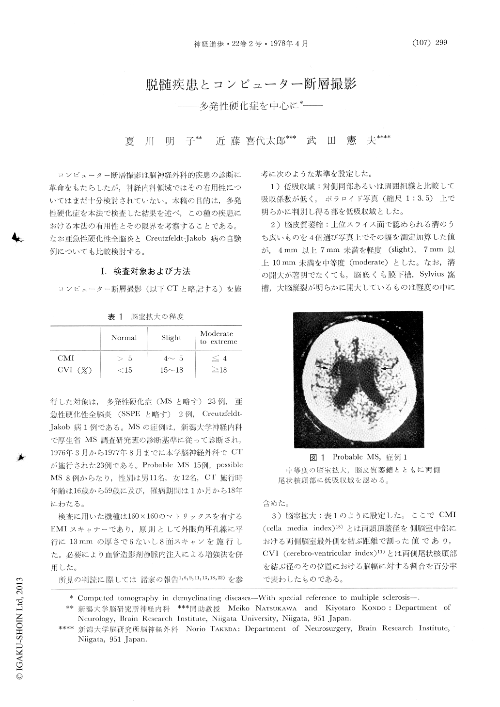
In 23 MS patients, 17 cases (73.9%) showed abnormal findings. Low density areas were seen in 4 of 23 (17.4%), cortical atrophy in 15 of 23 (65.2%), ventricular dilatation in 13 of 23 (56.5 %), brainstem atrophy in 4 of 22 (18.2%), and cerebellar atrophy in 1 of 22 cases (4.5%).
Low density areas, all except one, were located around the lateral ventricles and were presumed to represent MS plaques. Contrast enhancement was carried out in 3 cases without a positive result.

Copyright © 1978, Igaku-Shoin Ltd. All rights reserved.


