Japanese
English
- 有料閲覧
- Abstract 文献概要
- 1ページ目 Look Inside
I.はじめに
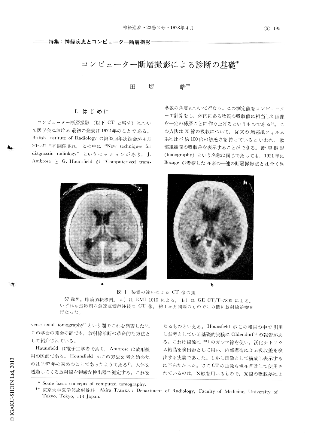
コンピューター断層撮影(以下CTと略す)について医学会における最初の発表は1972年のことである。British Institute of Radiologyの第32回年次総会が4月20〜21日に開催され,この中に"New techniques fordiagnostic radiology"というセッションがあり,J.AmbroseとG.Hounsfieldが"Computerized transverse axial tomography"という題でこれを発表した1)。この学会の開会の辞でも,放射線診断の革命的な方法として紹介されている。
Hounsfieldは電子工学者であり,Ambroseは放射線科の医師である。Hounsfieldがこの方法を考え始めたのは1967年の初めのことであったようである2)。人体を透過してくる放射線を鋭敏な検出器で測定する。これを多数の角度について行なう。この測定値をコンピューターで計算をし,体内にある物質の吸収値に相当した画像を一定の薄層ごとに作り上げるというものである3)。この方法はX線の吸収について,従来の増感紙フィルム系に比べ約100倍の敏感さを持っているといわれ,軟部組織間の吸収差を表示することができる。
Computed tomography has proven to be extremely useful for diagnosis in intracranial diseases. However, the successful use of this new technique depends on the appreciation of several basic features. The CT image represents a graphical display of computed numerical value and differs from conventional radiograms. Some important basic concepts; reconstructed picutre elements, section thickness, spatial resolution, CT numbers, viewing, artifacts and patient positioning are described and discussed.

Copyright © 1978, Igaku-Shoin Ltd. All rights reserved.


