Japanese
English
- 有料閲覧
- Abstract 文献概要
- 1ページ目 Look Inside
I.はじめに
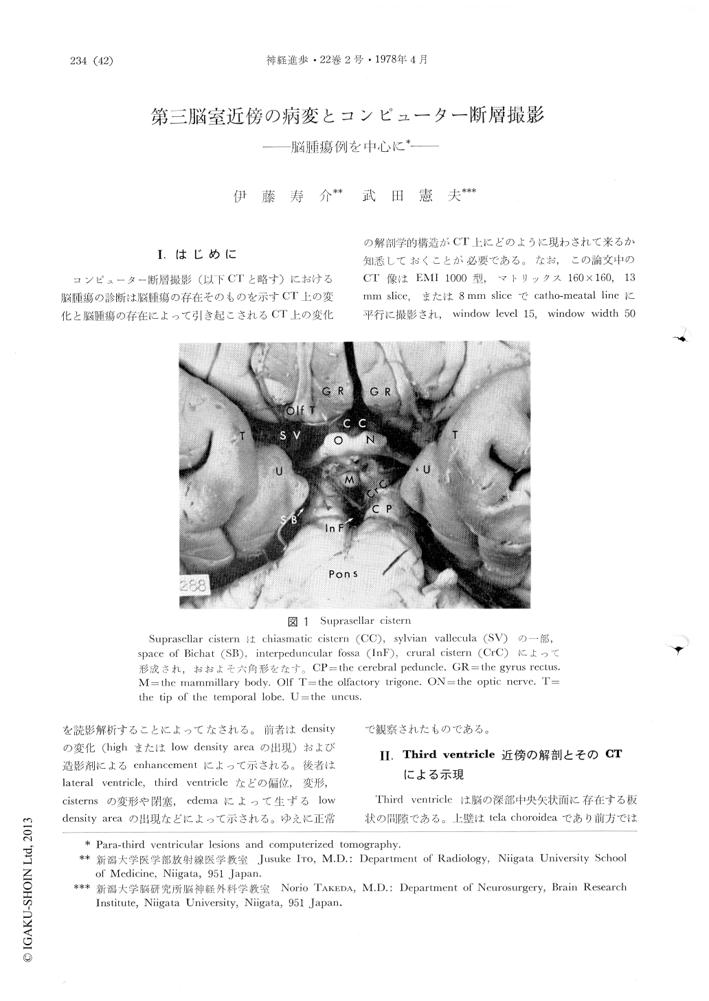
コンピューター断層撮影(以下CTと略す)における脳腫瘍の診断は脳腫瘍の存在そのものを示すCT上の変化と脳腫瘍の存在によって引き起こされるCT上の変化を読影解析することによってなされる。前者はdensityの変化(highまたはlow density areaの出現)および造影剤によるenhancementによって示される。後者ではlateral Ventricle,third ventricleなどの偏位,変形、cisternsの変形や閉塞,edemaによって生ずるlowdensity areaの出現などによって示される。ゆえに正常の解剖学的構造がCTで上にどのように現わされて来るか知悉しておくことが必要である。なお,この論文中のCT像はEMI1000型,マトリックス160×160,13mm slice,または8mm sliceでcathomeatal lineに平行に撮影され,window level 15,window width 50で観察されたものである。
Anatomy of the third ventricle and adjacent structures and their visualization by computerizedtomography (CT) have been described. CT findings of para-third ventricular tumors such as pituitary adenomas, craniopharyngiomas, tuberculum sellae meningioma, suprasellar germinoma, pineal region tumor, thalamic glioma, and some vascular lesions such as thalamic hemorrhage, aneurysm of the top of the basilar artery have been presented and discussed. Some difficulty in predicting histology of para-third ventricular tumors based on CT findings alone is stressed.

Copyright © 1978, Igaku-Shoin Ltd. All rights reserved.


