Japanese
English
- 有料閲覧
- Abstract 文献概要
- 1ページ目 Look Inside
I.緒言
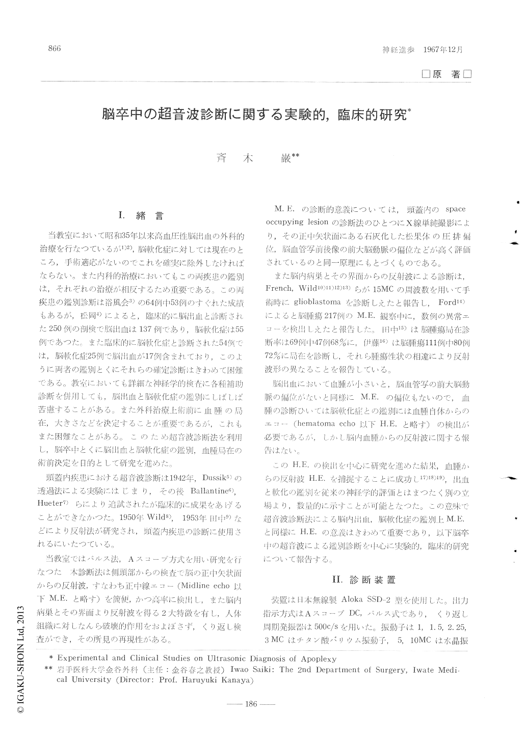
当教室において昭和35年以来高血圧性脳出血の外科的治療を行なつているが1)2),脳軟化症に対しては現在のところ,手術適応がないのでこれを確実に除外しなければならない。また内科的治療においてもこの両疾患の鑑別は,それぞれの治療が相反するため重要である。この両疾患の鑑別診断は浴風会3)の64例中53例のすぐれた成績もあるが,松岡4)によると,臨床的に脳出血と診断された250例の剖検で脳出血は137例であり,脳軟化症は55例であつた。また臨床的に脳軟化症と診断された54例では,脳軟化症25例で脳出血が17例含まれており,このように両者の鑑別とくにそれらの確定診断はきわめて困難である。教室においても詳細な神経学的検査に各種補助診断を併用しても,脳出血と脳軟化症の鑑別にしばしば苦慮することがある。また外科治療上術前に血腫の局在,大きさなどを決定することが重要であるが,これもまた困難なことがある。このため超音波診断法を利用し,脳卒中とくに脳出血と脳軟化症の鑑別,血腫局在の術前決定を目的として研究を進めた。
頭蓋内疾患における超音波診断は1942年,Dussik5)の透過法による実験にはじまり,その後Ballantine6),Hueter7)らにより追試されたが臨床的に成果をあげることができなかつた。1950年Wild8),1953年田中9)などにより反射法が研究され,頭蓋内疾患の診断に使用されるにいたつている。
Although a discrimination of apoplexy was, in the past, based on a clinical progress and fre-quency of symptoms, I developed a method of ultrasonic diagnosis in order to make it easier and surer and got the following restlts.
1) 1-lematoma-echoes were easy to be detected in the band of 2.25 MC, and multiple spike echoes were found from experimental intracere-bral hematoma, without any abnormal echo from brain softening.
2) Multiple spike hematoma-echoes were obse-rved through the operation of clinical cases and the examination from just outside of dura at autopsy. It was considered from experiment and operation that multiple spikes were grounded upon such factors as clotted blood mixed in fluid blood, destroyed fragments of brain and the rough surface between hematoma and brain.
3) Intracranial echoes were found in 100% from within a half of oval with the length of 7cm by 4cm right above the entrance of external auditory meatus, but I observed no echo from sinciput, vertex and occiput chiefly because of the thickness of skull.
4) Midline-echo deviated within 1mm from the center scale of echogram in 100 cases of normal adults, with the mean value of 0. 3±0.24 mm. A displacement exceeding 1mm was regarded as abnormal.
5) Of the 52 cases of supratentorial hemorrha-ge, hematoma-echoes were from the skin of head in 49 cases (94%) and the displacement of mi-dline-echo was in 43 (83 %). I could get the results of high rate in both.
6) As multiple spike echoes like hematoma-echoes were observed in no case and the displa-cement of midiline-echo was found in low rate in brain softening, the differentiation between the brain softening and intracerebral hemorrhage was possible in high rate. However, I could not differentiate brain softening from subarachnoideal hemorrhage.
Hematoma-echoes were observed in all of the 28 cases of supratentorial hemorrhage from just outside of dura during operation, which made a search for hematoma and operative technique easier.
From above mentioned studies, it was concl-tided that through the development of ultrasonic diagnosis in apoplexy it has become possible to differentiate intracerebral hemorrhage from brain softening with ease and in higher rate by means of quantitative judgment. It could also be said that owing to the easiness and quickness of the discrimination and the harmlessness to patients, this method was suitable for apoplexy in which many patients were in a serious condition, and should be put to practical use to a great extent in the future.

Copyright © 1967, Igaku-Shoin Ltd. All rights reserved.


