Japanese
English
- 有料閲覧
- Abstract 文献概要
- 1ページ目 Look Inside
I.はしがき
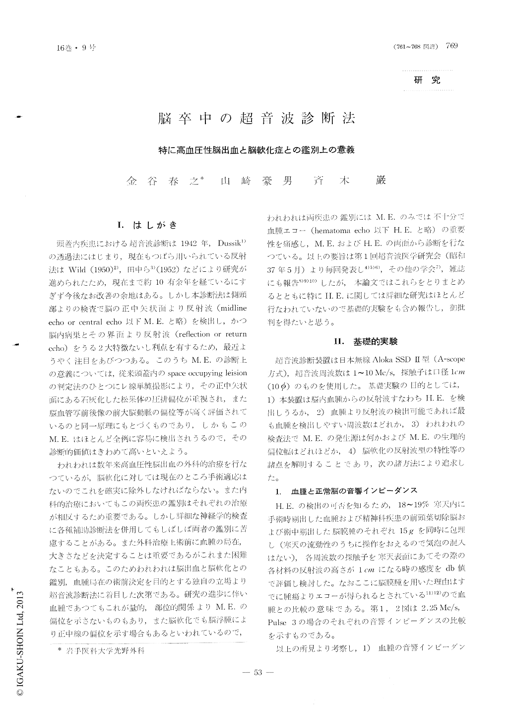
頭蓋内疾患における超音波診断は1942年,Dussik1)の透過法にはじまり,現在もつぱら用いられている反射法はWild (1950)2),田中ら3)(1952)などにより研究が進められたため,現在まで約10有余年を経ているにすぎず今後なお改善の余地はある。しかし本診断法は側頭部よりの検査で脳の正中矢状面より反射波(midline echo or central echo以下M. E.と略)を検出し,かつ脳内病巣とその界面より反射波(reflection or return echo)をうる2大特徴ないし利点を有するため,最近ようやく注目をあびつつある。このうちM. E.の診断上の意義については,従来頭蓋内のspace occupying leision の判定法のひとつにレ線単純撮影により,その正中矢状面にある石灰化した松果体の圧排偏位が重視され,また脳血管写前後像の前大脳動脈の偏位等が高く評価されているのと同一原理にもとづくものであり,しかもこのM. E.はほとんど全例に容易に検出されうるので,その診断的価値はきわめて高いといえよう。
われわれは数年来高血圧性脳出血の外科的治療を行なつているが,脳軟化に対しては現在のところ手術適応はないのでこれを確実に除外しなければならない。また内科的治療においてもこの両疾患の鑑別はそれぞれの治療が相反するため重要である。しかし詳細な神経学的検査に各種補助診断法を併用してもしばしば両者の鑑別に苦慮することがある。また外科治療上術前に血腫の局在,大きさなどを決定することは重要であるがこれまた困難なこともある。このためわれわれは脳出血と脳軟化との鑑別,血腫局在の術前決定を目的とする独自の立場より超音波診断法に着目した次第である。研究の進歩に伴い血腫であつてもこれが量的,部位的関係よりM. E.の偏位を示さないものもあり,また脳軟化でも脳浮腫により正中線の偏位を示す場合もあるといわれているので,われわれは両疾患の鑑別にはM. E.のみでは不十分で血腫エコー(hematoma echo以下H. E.と略)の重要性を痛感し,M. E.およびH. E.の両面から診断を行なつている。以上の要旨は第1回超音波医学研究会(昭和37年5月)より毎回発表し4)5)6),その他の学会7),雑誌にも報告8)9)10)したが,本論文ではこれらをとりまとめるとともに特にH. E.に関しては詳細な研究はほとんど行なわれていないので基礎的実験をも含め報告し,御批判を得たいと思う。
It is important from the point of surgical treat but very difficult sometimes, that intracerebral hemor-rhage is differentiated exactly from brain softening. As diagnosis of a small hematoma in brain can not be determined by only midline echo-shift in echogram, echo derived from hematoma itself were studied as a better method.
1) Multiple spikes were also seen in echogram of agar, in which clot were enclosed, though they were not seen in one of agar, in which specimen of meningioma or normal brain tissue were enclosed.
2) Multiple spikes were observed in echogram from autopsied brain of intracerebral hematoma.
3) Multiple spike echoes were observed in all cases of hypertensive intracerebral hemorrhage in echogram from just outside of dura during operation as same as in one from scalp.
4) Multiple spike echoes were found in 28 of 31 cases in the acute stage of intracerebral hemorrhage.
5) Midline-echo deviated within 1mm from the center scale of echogram in 100 cases of normal adults. Midline-echo shifted from normal range in echogram in 21 of 31 patients of acute hypertensive hemorrhage.
From the above mentioned study, it was concluded that the multiple spike echoes observed in echogram in cases of intracerebral hemorrhage were the echo derived from hematoma itself, and it was emphasized that these echoes were important for differentiation between hemorrhage and brain softening.

Copyright © 1964, Igaku-Shoin Ltd. All rights reserved.


